

Impact of Low Dose Oral Exposure to Bisphenol A (BPA) on the Neonatal Rat Hypothalamic and Hippocampal Transcriptome: A CLARITY-BPA Consortium Study
Sheryl E. Arambula, Scott M. Belcher, Antonio Planchart, Stephen D. Turner, and Heather B. Patisaul.
Endocrinology (2016).
DOI: https://doi.org/10.1210/en.2016-1339
PMID: 27571134
Publication
Abstract
Bisphenol A (BPA) is an endocrine disrupting, high volume production chemical found in a variety of products. Evidence of prenatal exposure has raised concerns that developmental BPA may disrupt sex-specific brain organization and, consequently, induce lasting changes on neurophysiology and behavior. We and others have shown that exposure to BPA at doses below the no-observed-adverse-effect level can disrupt the sex-specific expression of estrogen-responsive genes in the neonatal rat brain including estrogen receptors (ERs). The present studies, conducted as part of the Consortium Linking Academic and Regulatory Insights of BPA Toxicity program, expanded this work by examining the hippocampal and hypothalamic transcriptome on postnatal day 1 with the hypothesis that genes sensitive to estrogen and/or sexually dimorphic in expression would be altered by prenatal BPA exposure. NCTR Sprague-Dawley dams were gavaged from gestational day 6 until parturition with BPA (0-, 2.5-, 25-, 250-, 2500-, or 25 000-μg/kg body weight [bw]/d). Ethinyl estradiol was used as a reference estrogen (0.05- or 0.5-μg/kg bw/d). Postnatal day 1 brains were microdissected and gene expression was assessed with RNA-sequencing (0-, 2.5-, and 2500-μg/kg bw BPA groups only) and/or quantitative real-time PCR (all exposure groups). BPA-related transcriptional changes were mainly confined to the hypothalamus. Consistent with prior observations, BPA induced sex-specific effects on hypothalamic ERα and ERβ (Esr1 and Esr2) expression and hippocampal and hypothalamic oxytocin (Oxt) expression. These data demonstrate prenatal BPA exposure, even at doses below the current no-observed-adverse-effect level, can alter gene expression in the developing brain.
Figures
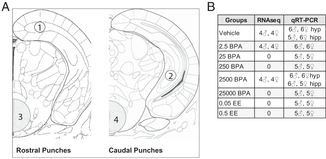
Figure 1. 2 hypothalamic punches and 4 hippocampal punches were obtained for each animal.
A, Anatomical representation of regions extracted via micropunch (obtained by approaching the regions of interest caudally and punching rostrally) and used for gene expression analysis. For each animal, 1 pair of bilateral anterodorsal hippocampal punches (1, unshaded) and 1 pair of bilateral caudoventral hippocampal punches (2, unshaded) were made, each 0.5 mm in diameter and 1.00 mm in depth. All 4 punches were combined, collectively comprising the whole hippocampus. Hypothalamic tissue consisted of 2 sequential punches (1.25 mm in diameter and 1.00 mm in depth): 1 anteromedial (3, shaded) and 1 caudomedial (4, shaded). B, Sample sizes for RNA-seq and qRT-PCR.
- Figure 1 (335 KB)
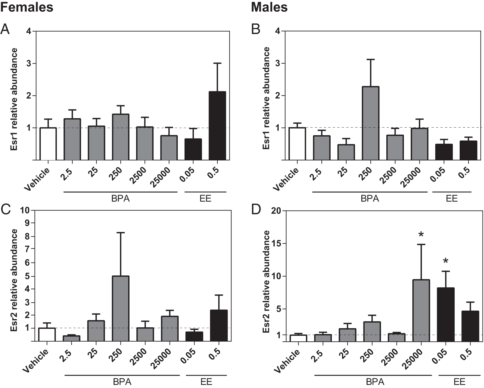
Figure 2. Effects of gestational BPA and EE on neonatal hippocampal ER expression.
BPA or EE did not affect Esr1 expression in females (A) or males (B).
In females, there was no effect of BPA or EE on Esr2 expression (C).
In males, exposure to the highest BPA dose and lowest EE dose resulted in significantly increased Esr2 expression (D).
Graphs depict mean ± SEM; *, P ≤ .05.
- Figure 2 (105 KB)
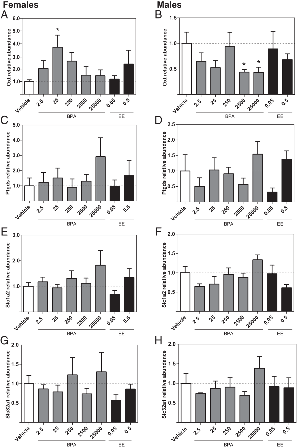
Figure 3. Effects of gestational BPA and EE exposure on neonatal hippocampal expression of selected genes.
In females, exposure to 25-μg BPA/kg bw/d significantly increased Oxt expression (A).
In males, exposure to 2500- and 25 000-μg BPA/kg bw/d decreased expression of Oxt (B).
BPA and EE had no significant effect on Ptgds (C and D), Slc1a2 (E and F), or Slc32a1 (G and H) expression in either females or males.
Graphs depict mean ± SEM; *, P ≤ .05.
- Figure 3 (235 KB)
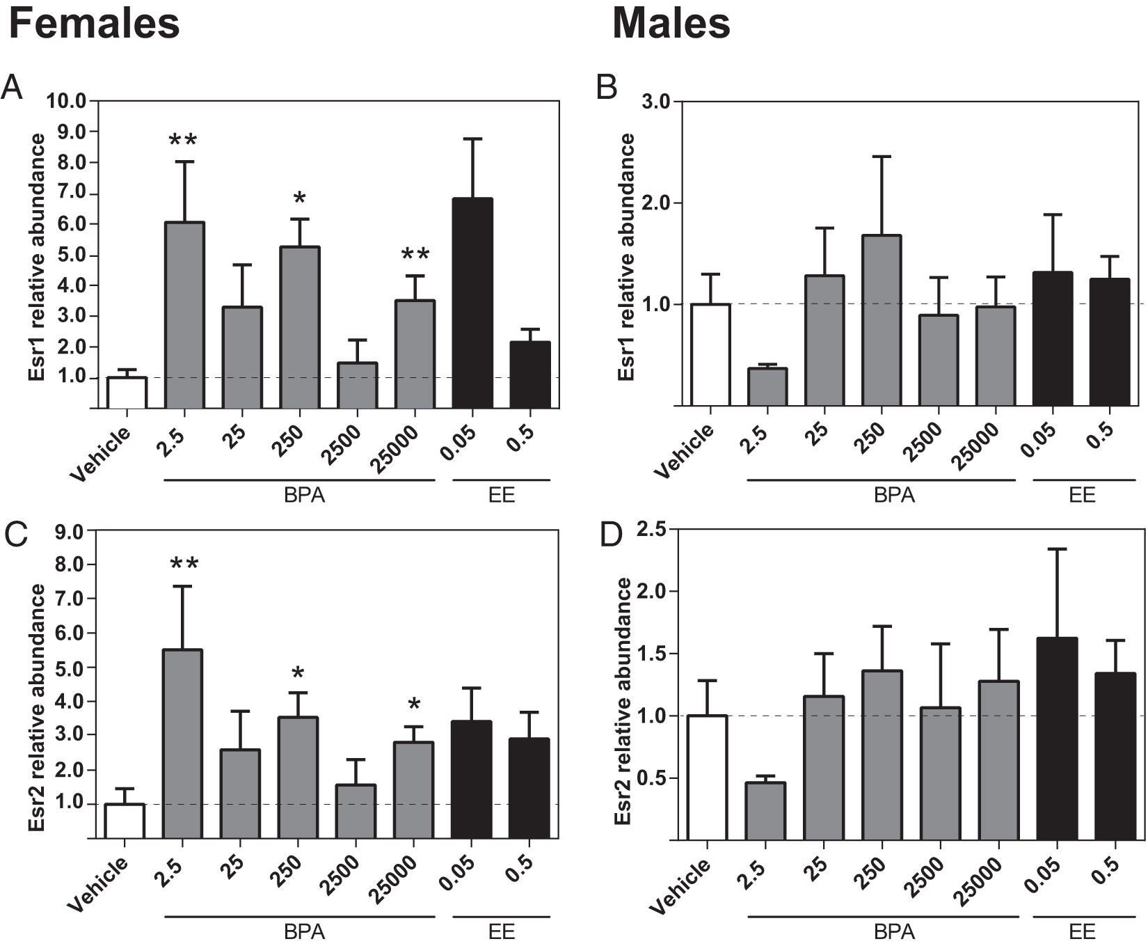
Figure 4. Effects of gestational BPA and EE exposure on neonatal hypothalamic ER expression.
Exposure to 2.5-, 250-, and 25 000-μg BPA/kg bw/d increased female expression of Esr1 and Esr2 (A and C).
Expression in males was unaffected (B and D).
EE had no effect on ER expression in either sex.
Graphs depict mean ± SEM; *, P ≤ .05 and **, P ≤ .01.
- Figure 4 (134 KB)
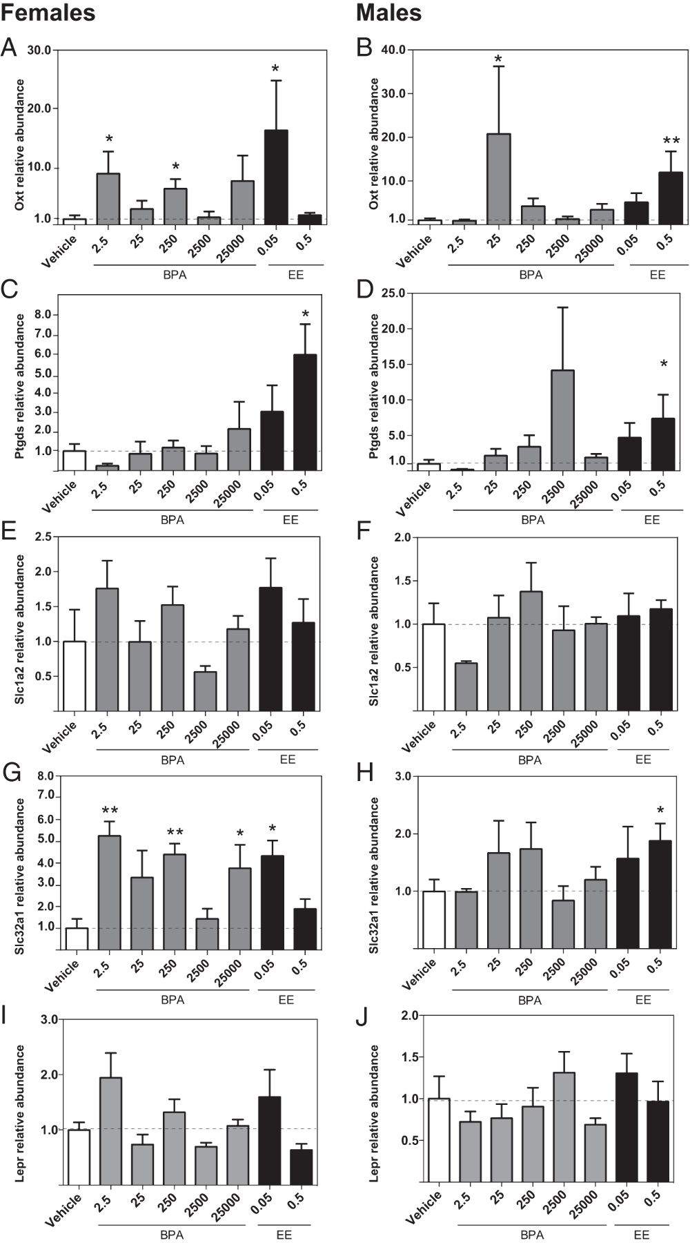
Figure 5. Effects of gestational BPA or EE on neonatal hypothalamic expression of selected genes.
Exposure to 2.5- and 250-μg BPA/kg bw/d and 0.05-μg EE/kg bw/d increased female expression of Oxt.
A, In males, 25-μg BPA/kg bw/d and the highest dose of EE increased Oxt expression (B).
Only the highest dose of EE affected Ptgds, increasing expression in both females and males (C and D).
Slc1a2 expression levels were not affected by BPA or EE in either sex (E and F).
The low dose of EE and 2.5-, 250-, and 25 000-μg BPA/kg bw/d masculinized (increased) female expression of Slc32a1 (G).
The high dose of EE increased male expression of Slc32a1 (H).
BPA or EE did not affect Lepr expression in either females (I) or males (J).
Graphs depict mean ± SEM; *, P ≤ .05 and **, P ≤ .01.
- Figure 5 (222 KB)
Figure 6. Sex differences in hippocampal and hypothalamic expression of selected genes.
Relative differences in gene expression between male and female control (unexposed) groups with male gene expression set as baseline.
A, No genes were sexually dimorphic in the neonatal hippocampus.
B, Esr2 and Slc32a1 exhibited male biased expression.
Graphs depict mean ± SEM; *, P ≤ .05 and **, P ≤ .01.
- Figure 6 (77 KB)
Tables
Table 1. Candidate and Novel Genes Assessed With qRT-PCR.
- Table 1 (278 KB)
Table 2. Differentially Expressed Genes for Which There Was a Significant Sex by Exposure Interaction at 2500-μg BPA/kg bw/d in Males.
- Table 2 (232 KB)
Table 3. qRT-PCR Outcomes and Descriptive Statistics for Genes Found to be Significantly Altered by BPA Exposure.
- Table 3 (113 KB)
Supplemental Materials
Supplementary Data
- Supplemental Table 1, 2, 4; Supplemental Figure 1 (4 MB)
- Supplemental Table 3: Overlapping significantly altered hypothalamic genes in males exposed to BPA. (80 KB)
- Supplemental Table 5: Compendium of results from the RNAseq and qPCR analyses for the genes of interest (Hippocampus, Hypothalmus). (38 KB)