

Identification of Vascular Disruptor Compounds by Analysis in Zebrafish Embryos and Mouse Embryonic Endothelial Cells
McCollum CW, Conde-Vancells J, Hans C, Vazquez-Chantada M, Kleinstreuer N, Tal T, Knudsen T, Shah SS, Merchant FA, Finnell RH, Gustafsson JÅ, Cabrera R, Bondesson M.
Reproductive Toxicology (2017)
DOI: https://doi.org/10.1016/j.reprotox.2016.11.005
PMID: 27838387
Publication
Abstract
To identify vascular disruptor compounds (VDCs), this study utilized an in vivo zebrafish embryo vascular model in conjunction with a mouse endothelial cell model to screen a subset of the U.S. Environmental Protection Agency (EPA) ToxCast Phase I chemical inventory. In zebrafish, 161 compounds were screened and 34 were identified by visual inspection as VDCs, of which 28 were confirmed as VDCs by quantitative image analysis. Testing of the zebrafish VDCs for their capacity to inhibit endothelial tube formation in the murine yolk-sac-derived endothelial cell line C166 identified 22 compounds that both disrupted zebrafish vascular development and murine endothelial in vitro tubulogenesis. Putative molecular targets for the VDCs were predicted using EPA's Toxicological Prioritization Index tool and a VDC signature based on a proposed adverse outcome pathway for developmental vascular toxicity. In conclusion, our screening approach identified 22 novel VDCs, some of which were active at nanomolar concentrations.
Figures
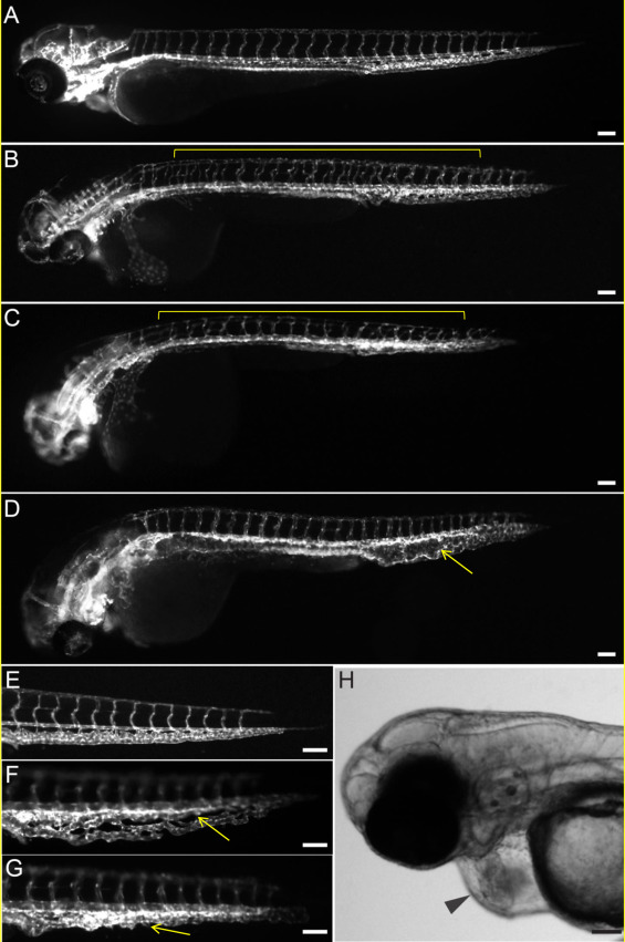
Figure 1. VDCs cause malformed vascular development in the ISV, DA and CVP.
Tg(kdrl:EGFP)mitfab692 embryos were treated with vehicle (control; A, E) or chemicals from the ToxCast Phase 1 chemical library (B-D, F, G) from 3 hpf to 72 hpf. Lateral view with anterior to the left and dorsal to the top. Vascular defects include non-overlapping ISVs (B), or thin and underdeveloped ISVs (C), expanded DA (D), and less condensed CVP (F), or misshapen CVP (G) and are marked with yellow lines or arrows. A common vascular-related phenotype, which is characteristic of cardiotoxicity, is pericardial edema (H; black arrowhead). Scale bar = 100 μm. (For interpretation of the references to colour in this figure legend, the reader is referred to the web version of this article.)
- Figure 1 (119 KB)
Figure 2. C166 cells tube formation is affected by VDCs.
C166 cells assemble into capillary-like structures when grown on Matrigel for 150 min. Angiogenesis analyzer mapping of the mesh network formed by C166 cells growing on Matrigel upon treatment with vehicle (0.1% DMSO) and increasing doses of rotenone for 150 min.
- Figure 2 (115 KB)
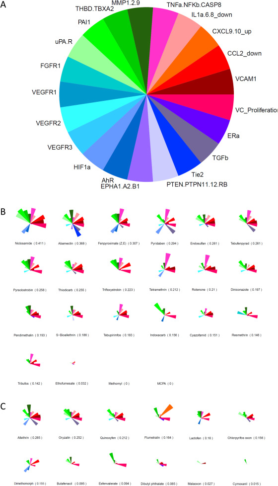
Figure 3. Color map showing minimal concentrations
The color map shows the minimal concentration of each chemical required to affect the parameters measured by angiogenesis analyzer in C166 cells.
- Figure 3 (83 KB)
Figure 4. pVDC signatures for zebrafish and C166 cell VDCs
Molecular pathways with corresponding in vitro assays used in the ToxCast screening program were selected to build the putative VDC signature (A). Ranking and pVDC signatures of VDCs active both in zebrafish and in C166 cells (B). Ranking and pVDC signatures of chemicals acting as VDCs in either zebrafish or C166 cells (C). Cytokine signaling (red); vessel stabilization (purple), angiogenic signaling (blue); and extracellular matrix (ECM) remodeling (green) quadrants are shown. (For interpretation of the references to colour in this figure legend, the reader is referred to the web version of this article.)
- Figure 4 (136 KB)
Tables
Table 1. Lowest effect levels (LELs) for compounds that cause ISV and PVC perturbations
* Compounds with “No effect” were initially found to cause vascular disruption via visual assessment, but not via automated image analysis. PE = Pericardial edema; BC = Blood clot, accumulation or hemorrhage; CVP = Caudal vein plexus phenotype; ISV = Intersegmental vessel phenotype; DA = Dorsal aorta; PCV = Posterior cardinal vein.
- Table 1 (12 KB)
Supplemental Materials
Supplemental Data
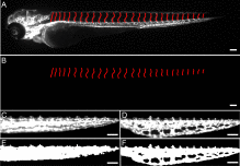
- Supplemental Figure S1: Perturbation of ISVs and CVP (19 KB)
- Supplemental Figure S2: The percentage of affected embryos (429 KB)
- Supplemental Figure S3: An example of data generated from the tube formation assay for all quantified parameters for rotenone (647 KB)
- Supplemental Table S1: List of chemicals tested (68 KB)
- Supplemental Table S2: Lowest effect levels (LELs) for compounds (11 KB)
- Supplemental Table S3: An example of data generated from the tube formation assay for all quantified parameters for rotenone (11 KB)