

Chemical Reactivity and Respiratory Toxicity of the α-Diketone Flavoring Agents 2,3-Butanedione, 2,3-Pentanedione, and 2,3-Hexanedione
Morgan DL, Jokinen MP, Johnson CL, Price HC, Gwinn WM, Bousquet RW, Flake GP.
Toxicol Pathol. 2016
DOI: https://doi.org/10.1177/0192623316638962
PMID: 27025954
Publication
Abstract
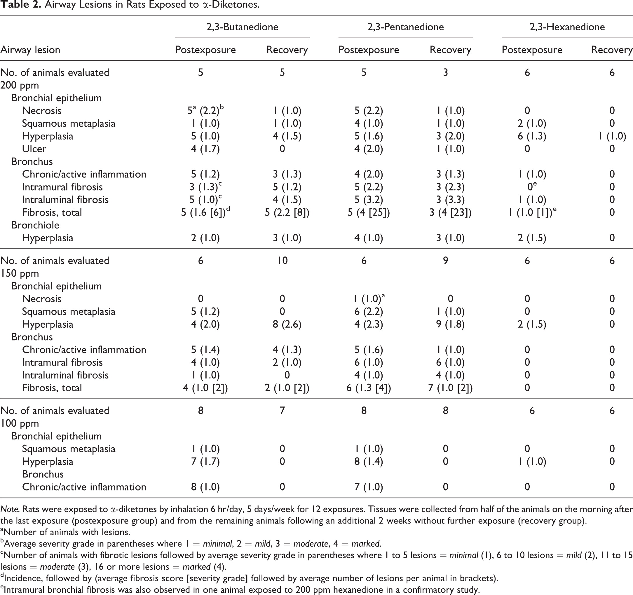
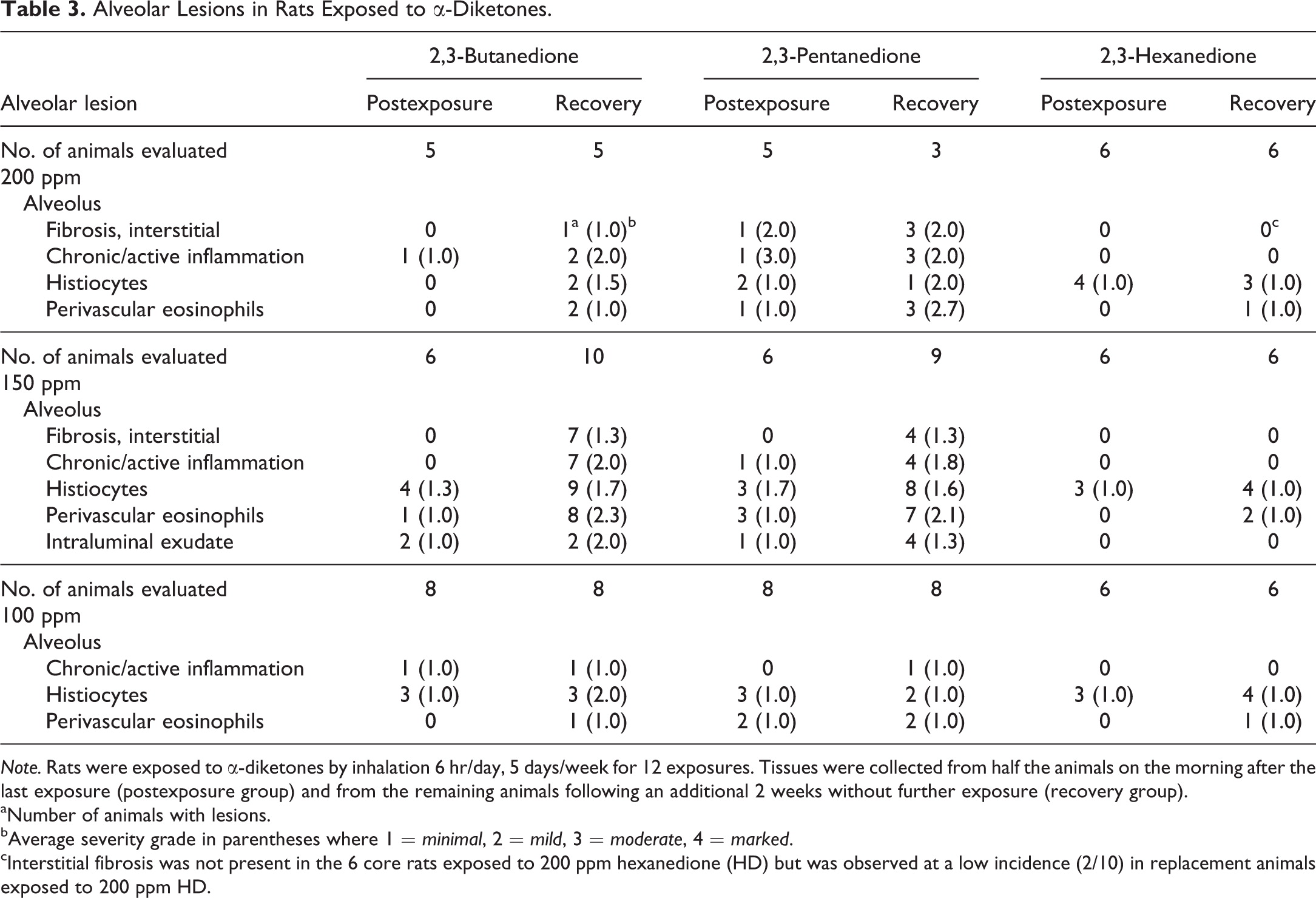
Occupational exposure to 2,3-butanedione (BD) vapors has been associated with severe respiratory disease leading to the use of potentially toxic substitutes. We compared the reactivity and respiratory toxicity of BD with that of two structurally related substitutes, 2,3-pentanedione (PD) and 2,3-hexanedione (HD). Chemical reactivity of the diketones with an arginine substrate decreased with increasing chain length (BD > PD > HD). Animals were evaluated the morning after a 2-week exposure to 0, 100, 150, or 200 ppm BD, PD, or HD (postexposure) or 2 weeks later (recovery). Bronchial fibrosis was observed in 5/5 BD and 5/5 PD rats at 200 ppm and in 4/6 BD and 6/6 PD rats at 150 ppm in the postexposure groups. Following recovery, bronchial fibrosis was observed in all surviving rats exposed to 200 ppm BD (5/5) or PD (3/3) and in 2/10 BD and 7/9 PD rats exposed to 150 ppm. Bronchial fibrosis was observed only in 2/12 HD-exposed rats in the 200 ppm postexposure group. Patchy interstitial fibrosis affected lungs of recovery groups exposed to 200 ppm PD (3/3) or BD (1/5) and to 150 ppm PD (4/9) or BD (7/10) and correlated with pulmonary function deficits. BD and PD were more reactive and produced more bronchial fibrosis than HD.
Figures
Figure 1. Body weights of rats exposed to diketones.
Body weights of rats exposed to 150 (▪) or 200 ppm (▴) of (A) 2,3-butanedione or (B) 2,3-pentanedione for 2 weeks were decreased relative to controls at all time points. Animals exposed to 200 ppm 2,3-butanedione or 2,3-pentanedione continued to lose weight during the 2-week recovery period. Exposure to 100 ppm (•) 2,3-butanedione or 2,3-pentanedione had no effect on body weights. (C) Exposure to 2,3-hexanedione did not cause significant changes in body weights, relative to air-exposed controls, at any time point.
- Figure 1 (1 MB)
Figure 2. Lung weights of rats exposed to diketones.
Absolute lung weights of animals exposed to (A) 2,3-butanedione, (C) 2,3-pentanedione, or (E) 2,3-hexanedione were not significantly different from controls in the postexposure groups (gray bars). Lung weights were significantly increased (p < .05) only in the 200 ppm 2,3-pentanedione recovery group (black bars; C). Relative lung weights (percentage of body weight) were significantly increased in postexposure and recovery groups of animals exposed to 150 or 200 ppm 2,3-butanedione (B) and were significantly increased in the postexposure 2,3-pentanedione groups exposed to 150 or 200 ppm, as well as in the 200 ppm recovery group (D). 2,3-Hexanedione had no significant effect on absolute or relative lung weights of postexposure or recovery animals (E, F). *p < .05; ***p < .001.
- Figure 2 (2 MB)
Figure 3. Pulmonary function of rats exposed to 150 ppm of each diketone.
No significant effects were detected in airway resistance or lung compliance in the postexposure groups exposed to 150 ppm 2,3-butanedione, 2,3-pentanedione, or 2,3-hexanedione (black bars) relative to controls (gray bars). However, after a 2-week recovery, airway resistance was significantly increased in 2,3-butanedione-exposed rats (p < .05), and compliance was significantly decreased in 2,3-butanedione- and 2,3-pentanedione-exposed rats (p < .05). No significant changes were observed in pulmonary function of 2,3-hexanedione-exposed rats in the postexposure or recovery groups. *p < .05.
- Figure 3 (2 MB)
Figure 4. Bronchial fibrotic lesions after 2 weeks of exposure to 200 ppm of each chemical.
(A) 2,3-Butanedione: intraluminal polyp, with a loose, fibromyxoid stroma, surrounded by low cuboidal regenerative epithelium. The polyp attached secondarily to the bronchial epithelium near its apical end (arrow). (B) 2,3-Butanedione: intramural fibrosis of a bronchial branch. The intima (asterisk) is uniformly thickened by loose, immature fibrous tissue, while the fibrosis of the adventitia (arrow) is dense. The bronchial epithelium has been replaced by a single cobblestone layer of regenerating cells with karyomegaly (arrowhead). (C) 2,3-Pentanedione: obliterative bronchitis, occluding the lumen of a large bronchus (arrowhead) and both branches (arrows). Mucus retention is present in the lumens of both branches. (D) 2,3-Pentanedione: obliterative bronchitis, small bronchial branch (arrow) just proximal to the preterminal bronchiole (not shown) and adjacent to an artery (arrowhead). The lumen is completely occluded by inflamed fibrous tissue containing a few small epithelial remnants. (E) 2,3-Hexanedione: small, intraluminal fibrotic bud (arrow), with mucosal epithelial hyperplasia at the base (arrowheads). (F) 2,3-Hexanedione: small focus of intramural loose fibroplasia (short arrow) and fibrosis (long arrow), with regenerating epithelium on the surface (arrowheads). These were the only 2 fibrotic lesions (E) and (F) identified in the bronchi with 2,3-hexanedione exposure. Original objective magnification: A = 20×, B = 20×, C = 4×, D = 4×, E = 10×, and F = 10×.
- Figure 4 (2 MB)
Figure 5. Bronchial lesions.
Bronchial lesions after 2 weeks of exposure to 200 ppm, followed by 2 weeks of recovery. (A) 2,3-Butanedione: intraluminal fibrosis with apparent luminal bridging by fibrotic buds (arrows) has focally obstructed the lumen of a secondary bronchus (asterisk) at the branch point from the main bronchial lumen (top, double asterisk). (B) 2,3-Butanedione: intramural fibrosis (asterisk), markedly thickening the submucosa of a large bronchus. (C) 2,3-Pentanedione: intraluminal fibrosis, with apparent luminal bridging by fibrotic polyps (center), partially obstructing the distal bronchial lumen (asterisk). The proximal bronchial lumen (double asterisk) is mucus filled due to fibrotic obstruction of the bronchial lumen upstream. (D) 2,3-Pentanedione: obliterative bronchiolitis. The lumen is almost completely occluded by dense fibro-collagenous tissue (arrowheads), with a few dispersed lymphocytes. (E) and (F) 2,3-Hexanedione: after the 2-week recovery period, no fibrotic bronchial lesions were found. The mucosal epithelium did exhibit areas of epithelial karyomegaly, focally with minimal hyperplasia (E, arrows), and areas of epithelial atrophy and/or regeneration (F, arrows). Original objective magnification: A = 4×, B = 10×, C = 2×, D = 10×, E = 20×, and F = 20×.
- Figure 5 (2 MB)
Figure 6. Obliterative bronchitis and obliterative bronchiolitis.
A, B, and C are deeper cuts of the 2-week lesion shown in Figure 4D. (A) Obliterative bronchitis. The lumen of this airway just proximal to the preterminal bronchiole is occluded by a loose, fibromyxoid tissue encircled by compressed remnants of squamous metaplastic mucosal epithelium (arrow). H&E. (B) Verhoeff van Gieson elastic stain reveals the delicate layer of elastica (arrow) between the epithelium and the muscularis. Note the thick elastic layer of the adjacent artery (arrowhead). (C) Masson trichrome stain demonstrates fibrotic thickening of the airway wall and adventitia, but little mature collagen within the fibromyxoid tissue of the occluded lumen (asterisk). D, E, and F are deeper cuts of the 4-week lesion shown in Figure 5D. (D) Obliterative bronchiolitis. The lumen of this terminal bronchiole is occluded by dense fibrous tissue, with only a remnant of the lumen remaining (arrow). H&E. (E) Verhoeff van Gieson elastic stain demonstrates a thin elastic layer (arrows) encircling the fibrotic lumen and remnant of mucosal epithelium, verifying the luminal occlusion. (F) Masson trichrome stain confirms the mature collagenous character of the fibrotic occlusion (arrow). Original objective magnification: A = 4×, B and C = 10×, D = 10×, E and F = 20×.
- Figure 6 (2 MB)
Figure 7. Bronchial intraluminal fibrosis, with luminal bridging.
Intraluminal fibrotic polyps often show foci of secondary attachment to the bronchial mucosal epithelium, especially in the 4 week animals (recovery group). (A) This is a deeper cut of the lesion shown in Figure 5C to illustrate that the intraluminal fibrotic process (arrows) does fully span the lumen, resulting in partial obstruction with accumulation of mucus in the lumen distally (asterisk). The accumulation of mucus on the proximal side of the fibrous bridge (double asterisk) is due to a second site of luminal fibrotic obstruction proximal to the one shown. (B) Immunohistochemical stain demonstrating expression of E-cadherin by the epithelial cells lining the fibrotic polyp (arrows) and the epithelial cells lining the bronchial lumens (arrowheads). Although the fibrotic bridge, and the epithelial cells and E-cadherins are continuous at this level, note that in Figure 5C only the epithelial cells are making contact. (C) and (D) These 2 fibrotic polyps each exhibit 4 separate sites of attachment to the bronchial mucosal epithelium (short arrows). The broader base, and presumably the origin, of each polyp is indicated by the long arrows. Note that there are also 2 additional foci of early epithelial bridging or attraction in C (arrowheads) and a slender epithelial bridge in D (arrowhead). We hypothesize that these foci of secondary attachment are due to the attraction of E-cadherins between the epithelial cells lining the polyps and the epithelial cells lining the bronchial lumens. Original object magnification: A=2x, B, C, and D=4x.
- Figure 7 (1 MB)
Figure 8. Pulmonary interstitial fibrosis.
Pulmonary interstitial fibrosis after 2 weeks of chemical exposure, followed by 2 weeks of recovery. (A) and (B) 2,3-Butanedione, 150 ppm: the alveolar walls in this area were thickened by interstitial fibrosis, which was centered around alveolar ducts, and accompanied by a chronic active inflammatory infiltrate. (C) and (D) 2,3-Pentanedione, 200 ppm: fibrosis was extensive in this lobe, predominantly subpleural, and relatively confluent in some areas. (E) and (F) 2,3-Hexanedione, 200 ppm: patchy, minimal interstitial fibrosis was identified in only 2 rats exposed to hexanedione and was characterized by prominent thickening of alveolar duct walls. Original objective magnification: A, C, and E = 4×; B, D, and F = 10×.
- Figure 8 (3 MB)
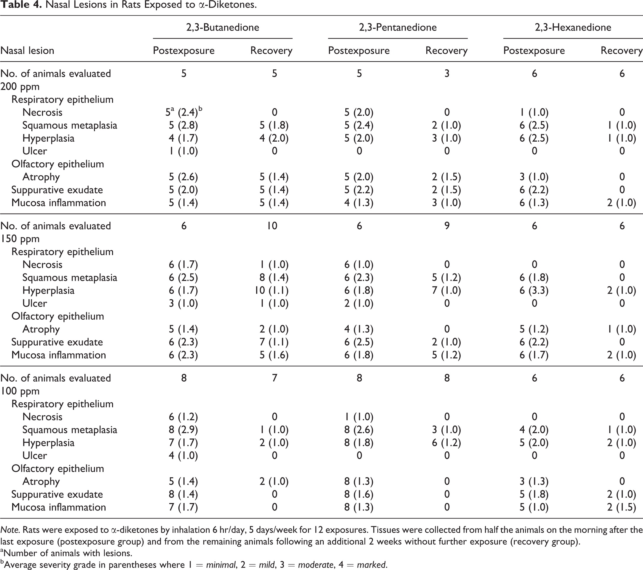
Figure 9. Nasal cavity lesions after 2 weeks of exposure to 200 ppm of each diketone.
Left half of panel shows nasoturbinate, level 1, from (A) 2,3-butanedione, (C) 2,3-pentanedione, and (E) 2,3-hexanedione. Exposure to each chemical resulted in inflammation, exudate, and squamous metaplasia (arrowheads). Respiratory epithelial necrosis (arrows), however, was common with 2,3-butanedione (A) and 2,3-pentanedione (C) but was noted focally in only one animal exposed to hexanedione. Right half of panel shows ethmoid turbinates, level 3, from (B) 2,3-butanedione, (D) 2,3-pentanedione, and (F) 2,3-hexanedione. Exposure to each chemical resulted in areas of olfactory epithelial atrophy (arrowheads), which can be better appreciated by comparison to the more normal olfactory epithelium on the opposite side of the turbinates (arrows). Note that the underlying nerve fibers and Bowman’s glands in the lamina propria are also atrophic (asterisks) beneath areas of epithelial atrophy (compare to the nerve fibers and glands beneath the more normal olfactory epithelium). Inflammatory exudate is present in the lumens between the turbinates. Original objective magnification for all photos: 10×.
- Figure 9 (2 MB)
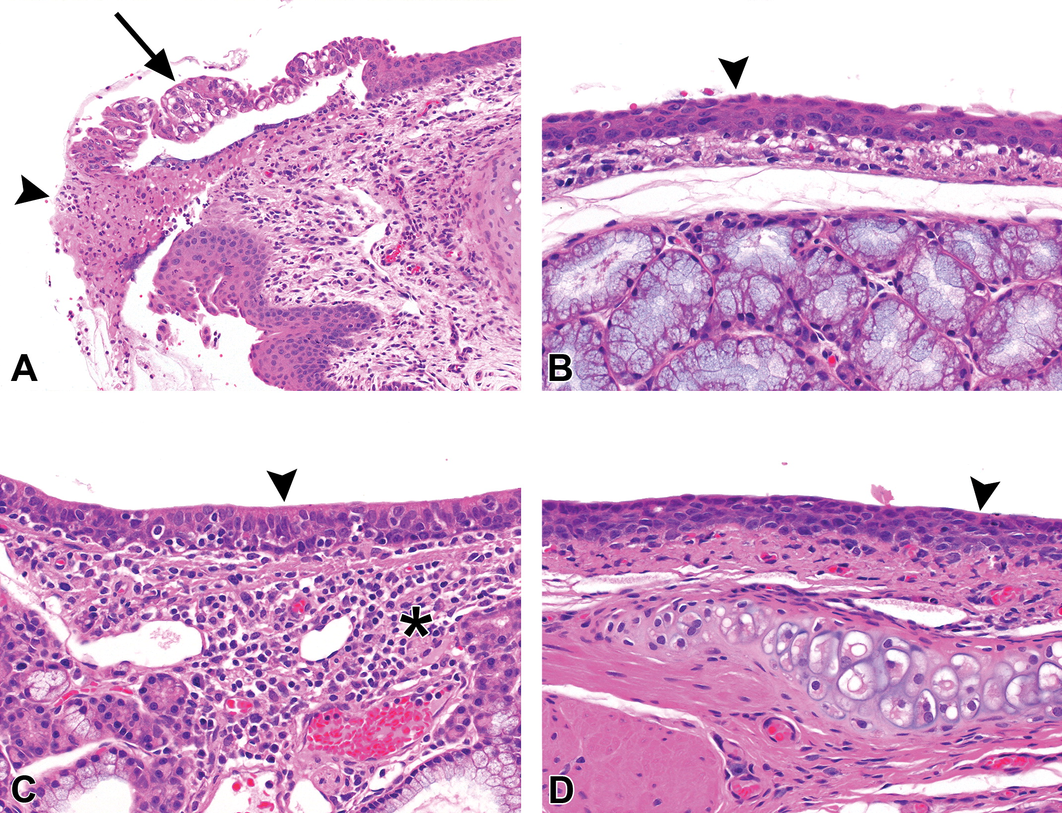
Figure 10. 2,3-Hexanedione: larynx and trachea.
2,3-Hexanedione: larynx and trachea after 2 weeks of exposure to 200 ppm (postexposure group). (A) Larynx: focal necrosis (arrowhead) of the epithelium overlying the arytenoid cartilage of level 1. The epithelium on the medial side of the necrosis was partially detached and degenerated (arrow). (B) Larynx: the respiratory epithelium at the base of the epiglottis has been replaced by squamous metaplastic epithelium (arrowhead). (C) Trachea: the submucosa in this rat contained a chronic inflammatory infiltrate (asterisk), and the epithelium was mildly hyperplastic (arrowhead). (D) Trachea: squamous metaplastic epithelium (arrowhead) has replaced the normal respiratory epithelium. Original object magnification: A=10x B, C, and D=20x.
- Figure 10 (1 MB)
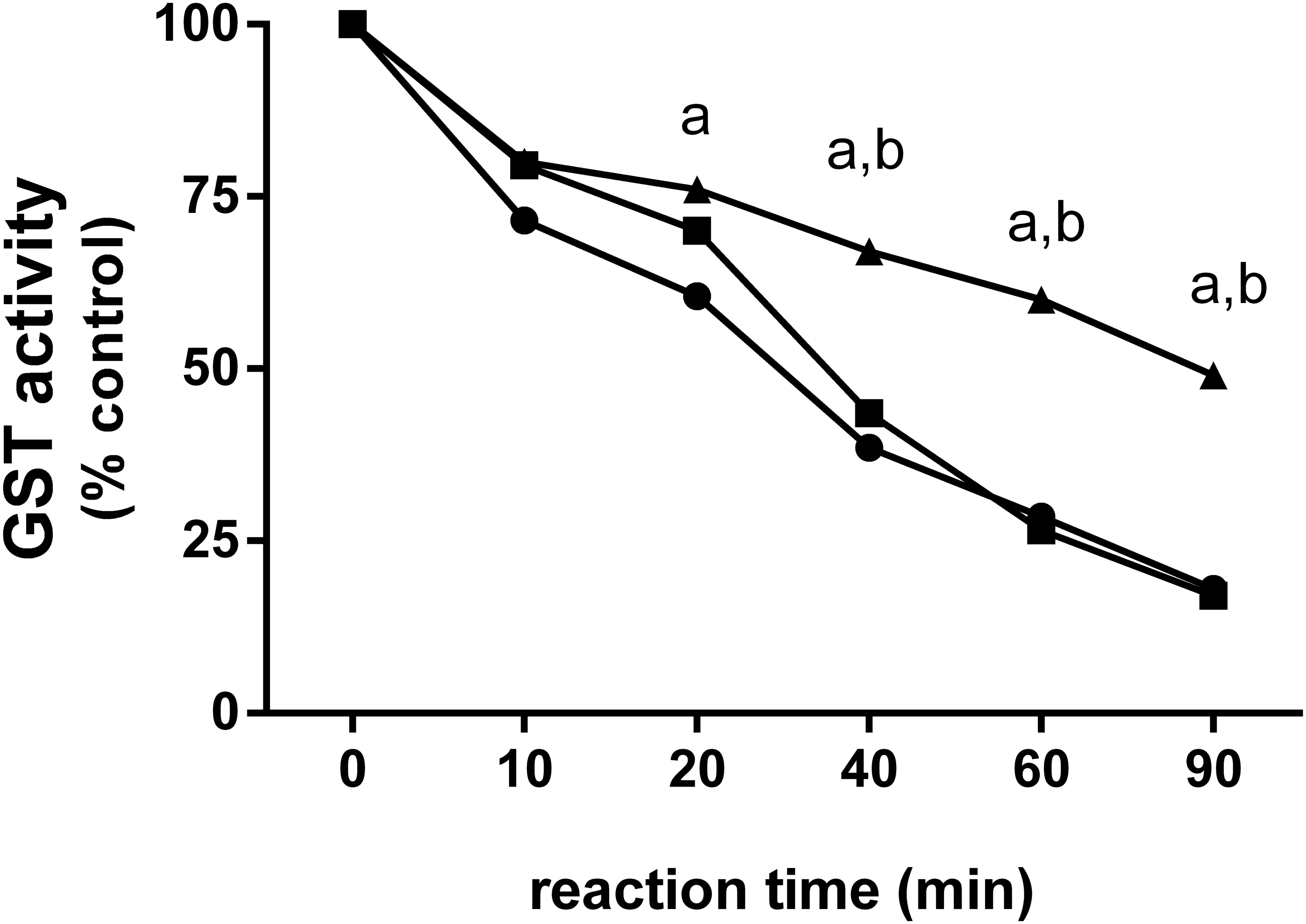
Figure 11. Relative reactivity of α-diketones with glutathione-S-transferase (GST).
2,3-Butanedione (•) and 2,3-pentanedione (▪) caused a relatively rapid inactivation of GST in a cell-free solution. 2,3-Butanedione appeared to have slightly greater reactivity with GST than 2,3-pentanedione initially, but these differences were not significantly different. 2,3-Hexanedione (▴) inactivated GST at a slower rate than 2,3-butanedione and 2,3-pentanedione, suggesting that 2,3-hexanedione is the least reactive; “a” indicates GST inactivation by hexanedione (HD) was significantly less than inactivation by 2,3-butanedione (p < .05); “b” indicates the GST inactivation by HD was significantly less than inactivation by 2,3-pentanedione (p < .05).
- Figure 11 (449 KB)
Tables
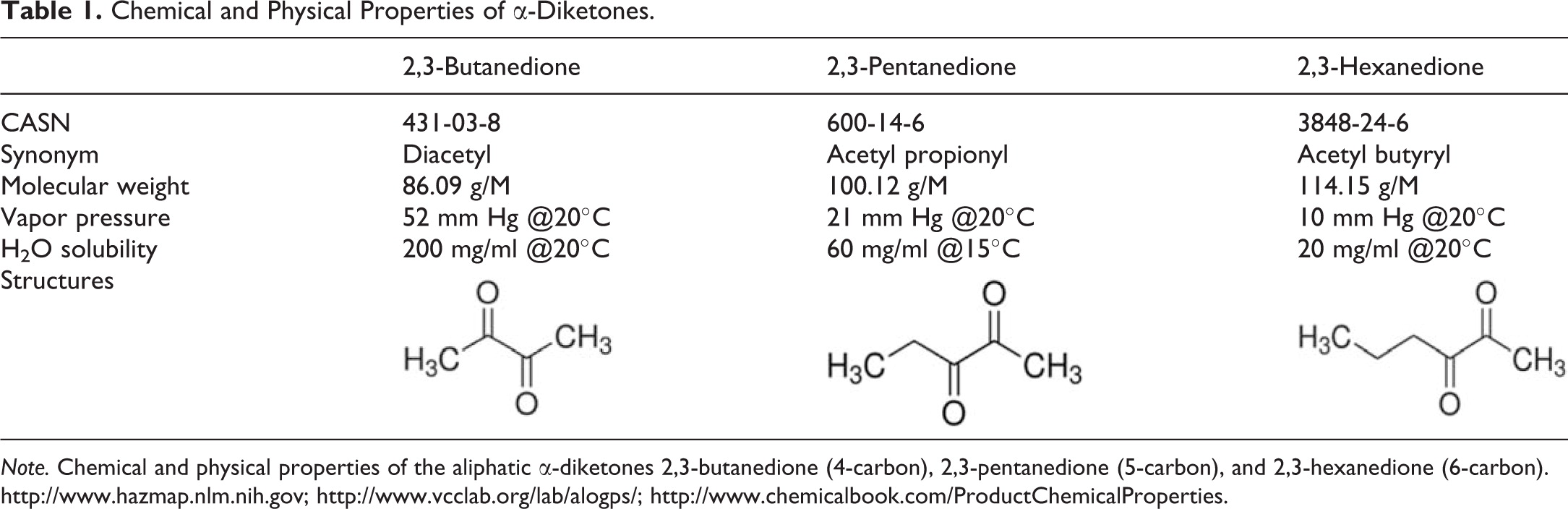
Table 1. Chemical and Physical Properties of α-Diketones
- Table 1 (244 KB)
Table 2. Airway Lesions in Rats Exposed to α-Diketones
- Table 2 (647 KB)
Table 3. Alveolar Lesions in Rats Exposed to α-Diketones
- Table 3 (484 KB)
Table 4. Nasal Lesions in Rats Exposed to α-Diketones
- Table 4 (582 KB)