

Evaluation of the Respiratory Tract Toxicity of ortho-Phthalaldehyde, a Proposed Alternative for the Chemical Disinfectant Glutaraldehyde
Natasha R. Catlin, Cynthia J. Willson, Matthew Stout, Grace E. Kissling, Suramya Waidyanatha, Gregory L. Baker, Barry K. Hayden, Michael Wyde.
Inhalation Toxicology (2017).
DOI: https://doi.org/10.1080/08958378.2017.1390015
PMID: 29039228
Publication
Abstract
ortho-Phthalaldehyde (OPA) is a high-level chemical disinfectant that is commonly used for chemical sterilization of dental and medical instruments as an alternative to glutaraldehyde, a known skin and respiratory sensitizer. Concern for safe levels of human exposure remains due to a lack of toxicity data as well as human case reports of skin and respiratory sensitization following OPA exposure. The present study evaluated the inhalational toxicity of OPA in Harlan Sprague–Dawley rats and B6C3F1/N mice. Groups of 10 male and female rats and mice were exposed to OPA by whole-body inhalation for 3 months at concentrations of 0 (control), 0.44, 0.88, 1.75, 3.5, or 7.0 ppm. Rats and mice developed a spectrum of lesions at sites of contact throughout the respiratory tract (nose, larynx, trachea, lung), as well as in the skin and eye, consistent with a severe irritant response. In general, histologic lesions (necrosis, inflammation, regeneration, hyperplasia and metaplasia) occurred at deeper sites within the respiratory tract with increasing exposure concentration. As a first site of contact, the nose exhibited the greatest response to OPA exposure and resulted in an increased incidence, severity and variety of lesions compared to a previous study of glutaraldehyde exposure at similar exposure concentrations. This increased response in the nasal cavity, combined with extensive lesions throughout the respiratory tract, provides concern for use of OPA as a replacement for glutaraldehyde as a high-level disinfectant.
Figures
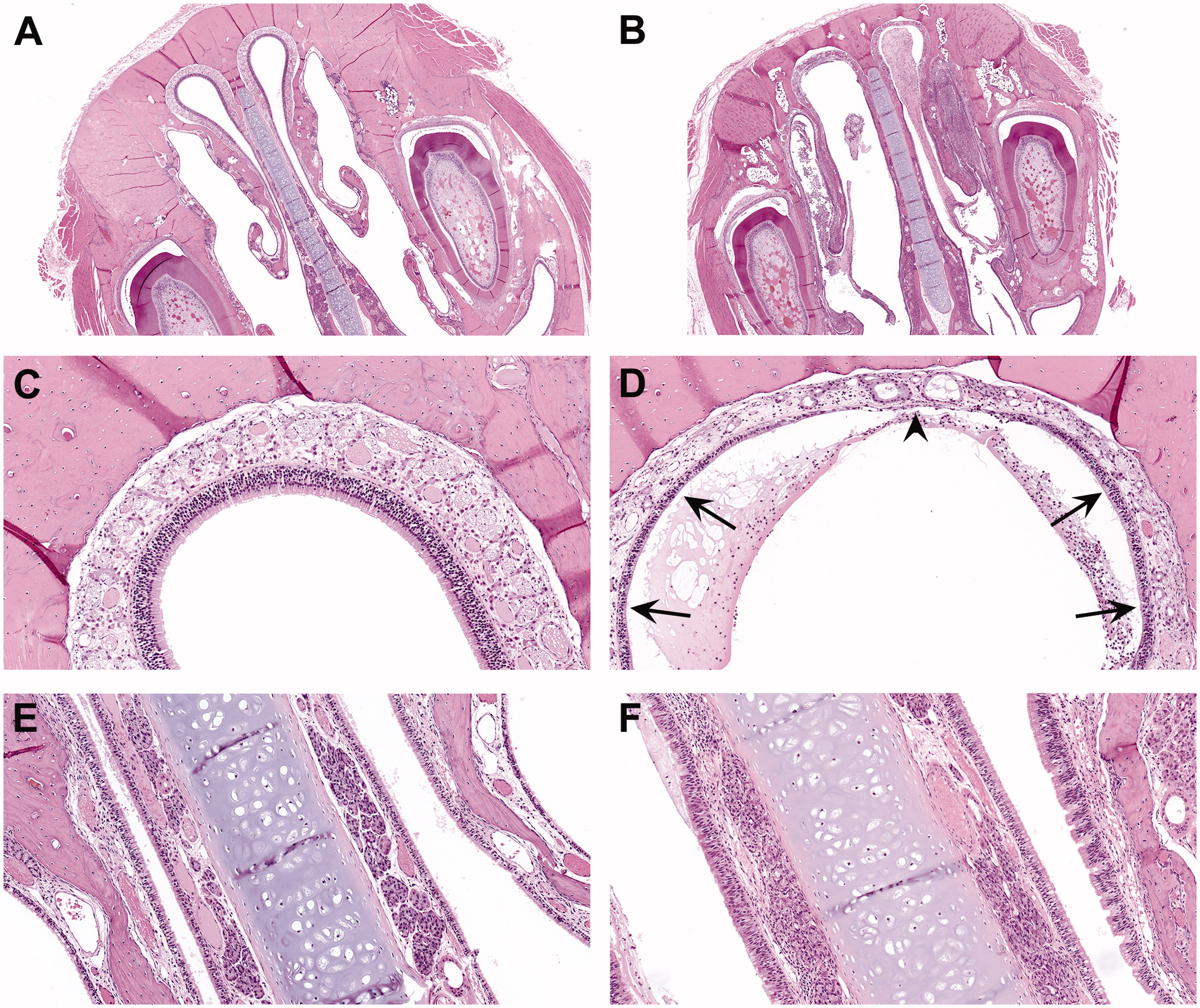
Figure 1. Representative microscopic lesions in the nose of Sprague–Dawley rats.
Representative microscopic lesions in the nose of Sprague–Dawley rats exposed to o-phthalaldehyde in the 3-month inhalation study.
(A) Normal Level II section of the nose from a control male rat. 2x.
(B) Suppurative inflammation and nasal turbinate atrophy in a Level II section of the nose from a male rat exposed to 3.5 ppm. Exudate containing degenerate neutrophils fills the lumen, and the nasoturbinates are distorted and blunted. Also, the olfactory epithelium lining the dorsal meatus is lost (necrosis). 2x.
(C) Normal olfactory epithelium in the dorsal meatus of a Level II section of the nose from a control male rat. 20x.
(D) Olfactory epithelium necrosis (arrowhead) and thinning (atrophy; arrows) in the dorsal meatus of a Level II section of the nose from a male rat exposed to 1.75 ppm. 20x.
(E) Normal respiratory epithelium along the nasal septum and nasoturbinates in a Level II section of the nose from a control male rat. 20x.
(F) The respiratory epithelium is multifocally thickened with nuclear crowding (respiratory epithelium hyperplasia), especially along the left side of the nasal septum and along the nasoturbinate in a Level II section of the nose from a male rat exposed to 0.88 ppm. 20x. H&E.
- Figure 1 (3 MB)
Figure 2. Representative microscopic lesions in the larynx, trachea, and lung of Sprague–Dawley rats
Representative microscopic lesions in the larynx, trachea, and lung of Sprague–Dawley rats exposed to o-phthalaldehyde in the 3-month inhalation study.
(A) Normal larynx at the base of the epiglottis from a control female rat. 20x.
(B) Squamous metaplasia of the larynx, seen as replacement of the normal respiratory epithelium of the larynx with multiple thickened layers of cuboidal to flattened epithelium, with keratinization along the luminal surface. 20x.
(C) Normal trachea from a control female rat. 4x.
(D) Necrosis and fibrosis in the trachea from a male rat exposed to 3.5 ppm. There is loss of the respiratory epithelium and an increase in fibrous connective tissue within the lamina propria of the trachea that partially occludes the tracheal lumen. 4x.
(E) Necrosis, inflammation, and fibrosis in the lung from a female rat exposed to 7.0 ppm. There is loss of the bronchial epithelium, and inflammatory cells within necrotic debris and mucus in the bronchi and within the peribronchial connective tissue. Bronchus fibrosis included both intraluminal and intramural changes. Intraluminal fibrosis included large inflammatory fibrotic polyploid structures extending into and partially occluding the bronchial lumen, whereas intramural fibrosis was thickening of the bronchial wall by similar connective tissue without projection into the lumen. 4x.
(F) Bronchial fibrosis (intraluminal and intramural) and inflammation in the lung from another female rat exposed to 7.0 ppm. 10x. H&E.
- Figure 2 (4 MB)
Tables
Table 1. Survival and relative final bodyweights of rats and mice.
Survival and relative final bodyweights of rats and mice in the 3-month inhalation studies of ortho-phthalaldehyde.
- Table 1 (149 KB)
Table 2. Incidences of respiratory lesions in male rats.
Incidences of respiratory lesions in male rats exposed to ortho-phthalaldehyde for 3 months.
- Table 2 (356 KB)
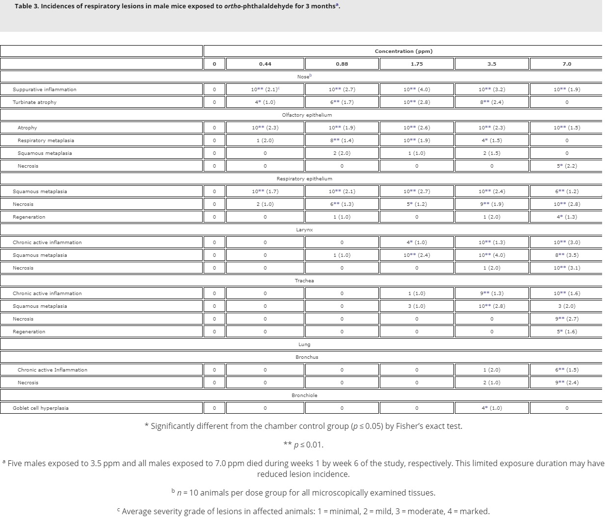
Table 3. Incidences of respiratory lesions in male mice.
Incidences of respiratory lesions in male mice exposed to ortho-phthalaldehyde for 3 months.
- Table 3 (294 KB)
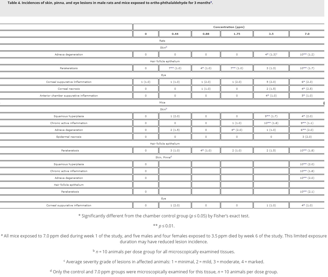
Table 4. Incidences of skin, pinna, and eye lesions in male rats and mice.
Incidences of skin, pinna, and eye lesions in male rats and mice exposed to ortho-phthalaldehyde for 3 months.
- Table 4 (282 KB)
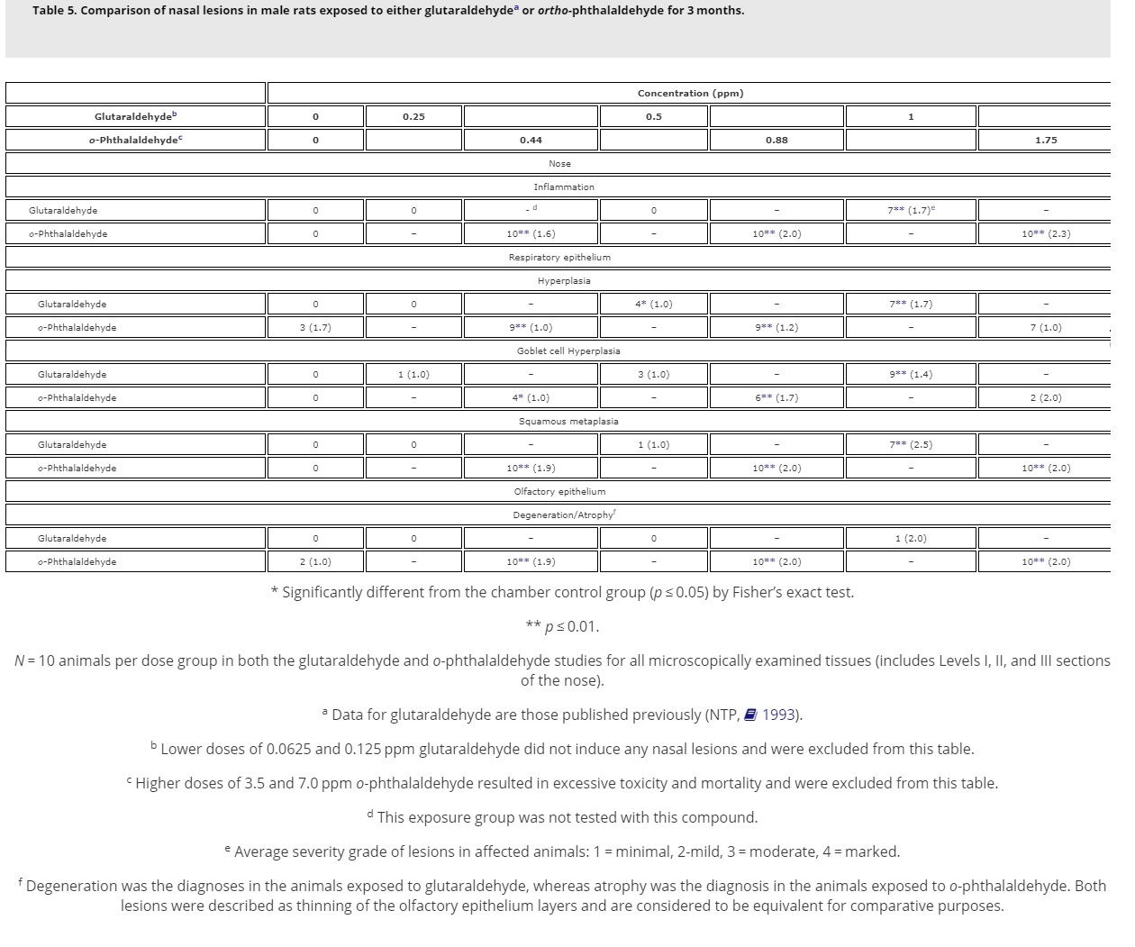
Table 5. Comparison of nasal lesions in male rats.
Comparison of nasal lesions in male rats exposed to either glutaraldehydea or ortho-phthalaldehyde for 3 months.
- Table 5 (266 KB)
Table 6. Comparison of nasal and laryngeal lesions in male mice.
Comparison of nasal and laryngeal lesions in male mice exposed to either glutaraldehydea or o-phthalaldehyde for 3 months.
- Table 6 (233 KB)