

Immunohistochemical Analysis of Rat Renal Tumours Caused by Ochratoxin A
Diana Herman and Peter Mantle.
Toxins (2017).
DOI: https://doi.org/10.3390/toxins9120384
PMID: 29182526
Publication
Abstract
Experimental renal cancer caused by ochratoxin A (OTA) in rats was first defined in the US National Toxicology Program (1989) and raised questions about any aetiological role in human urinary tract tumours. A review of histopathology in several rat kidney tumours from dietary OTA in recently described London studies, augmented by clinical immunohistochemistry for the first time for this mycotoxin, establishes their renal tubular cell origin. It had been assumed that the toxin might cause the human urothelial tumours associated with Balkan endemic nephropathy, but the present study could not support this. Comparison with a similar review of a metastasising renal tumour from a female rat of the NTP study consistently shows the kidney as the primary carcinogenic site for OTA. Morphological heterogeneity of these kidney tumours as epithelioid and/or sarcomatoid is revealed. Leiomyosarcoma was also diagnosed, and rhabdomyosarcoma differentiation was observed in the exceptionally aggressive NTP female tumour. The present pilot study involving immunohistochemistry indicates need for wider review of archived tumours for experimental evidence before formulating any epidemiological basis from a rat model for OTA’s relevance to idiopathic human renal cell carcinoma. Although the NTP study concluded that females are less sensitive to OTA than males, some female tumours still had heterogeneous morphology.
Figures
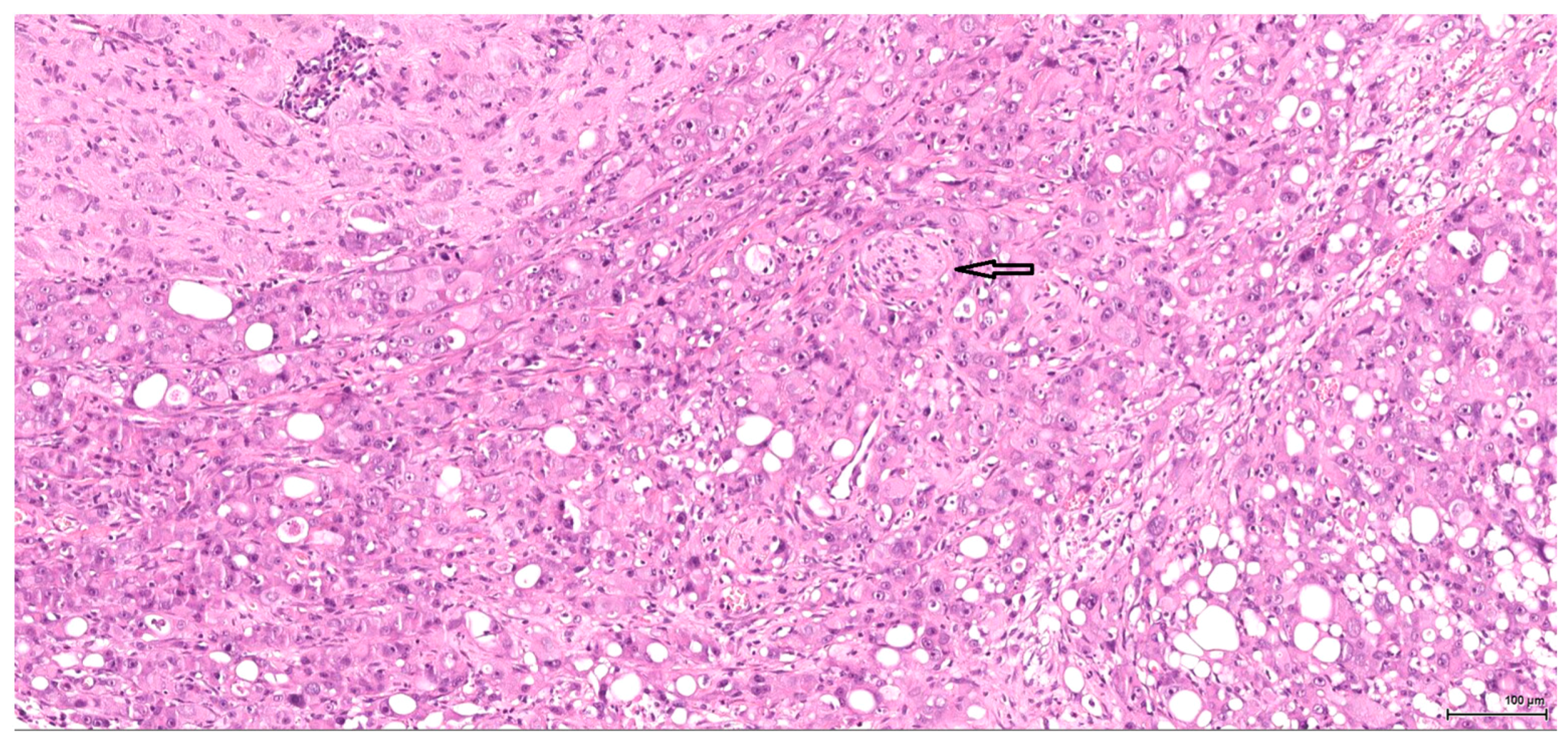
Figure 1. H&E: Male (case 1).
Epithelioid morphology, very focal clear cytoplasm, comedo-like tumour necrosis (arrows), pushing-type invasion front.
- Figure 1 (4 MB)
Figure 2. H&E: Male (case 4).
High-grade tumour with aggressive behaviour, infiltrating the perirenal fat (bottom right), nerves (arrow) and part of an autonomic ganglion (top left).
- Figure 2 (4 MB)
Figure 3. H&E: Male (case 3).
Marked intraepithelial cytological atypia within an enlarged tubule (arrow), adjacent to invasive tumour (right).
- Figure 3 (4 MB)
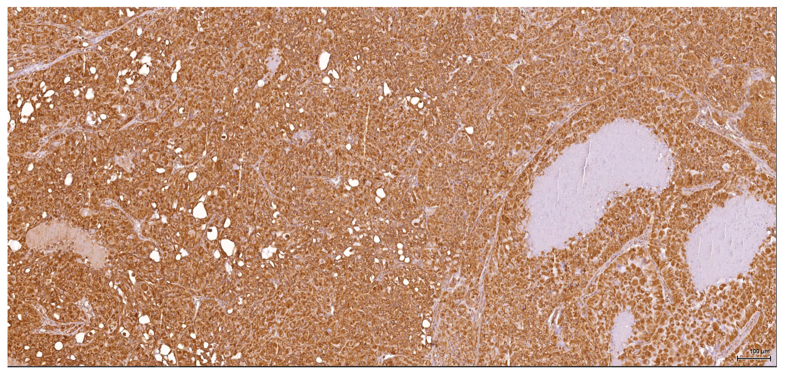
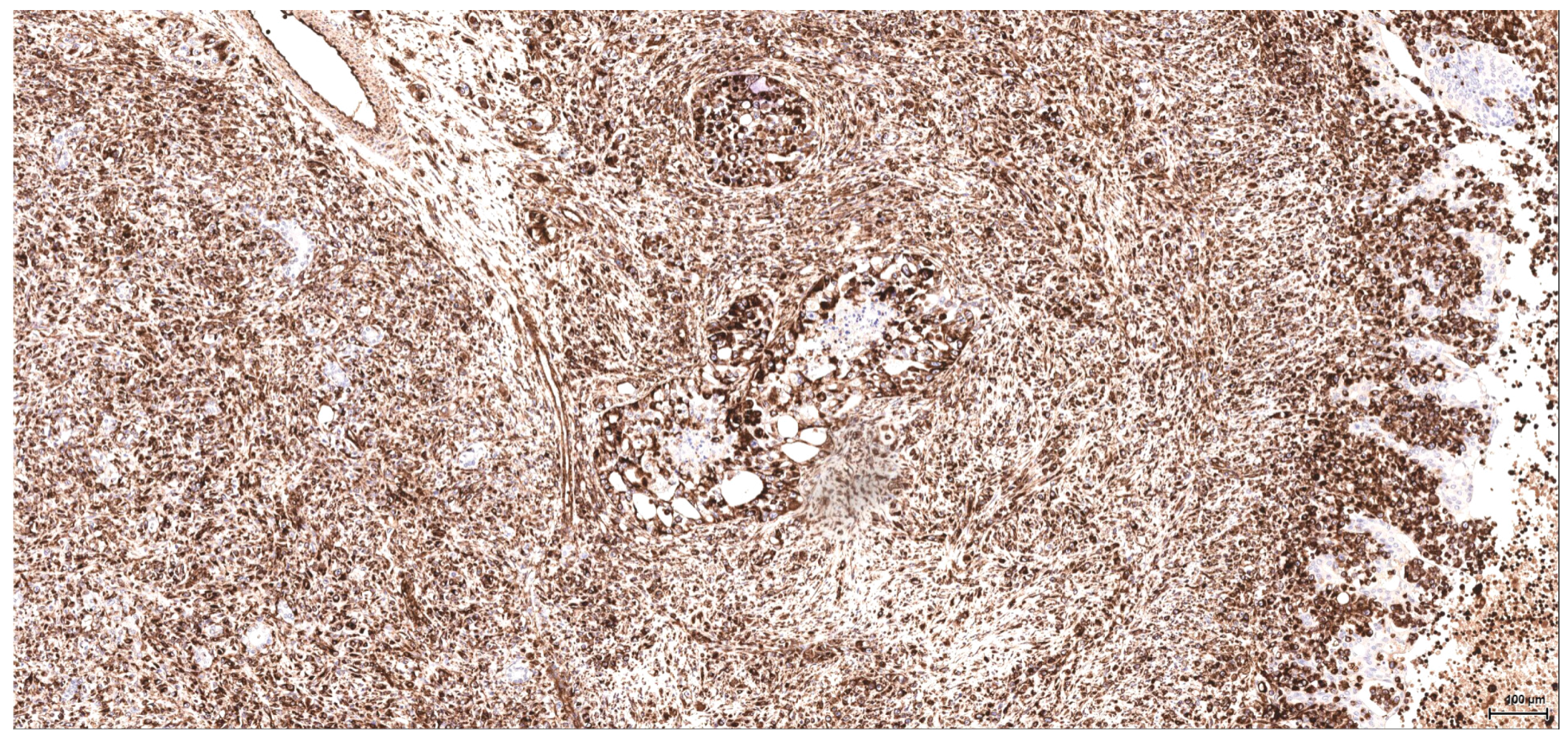
Figure 4. Vimentin: Male (case 2): intense and diffuse reaction.
- Figure 4 (5 MB)
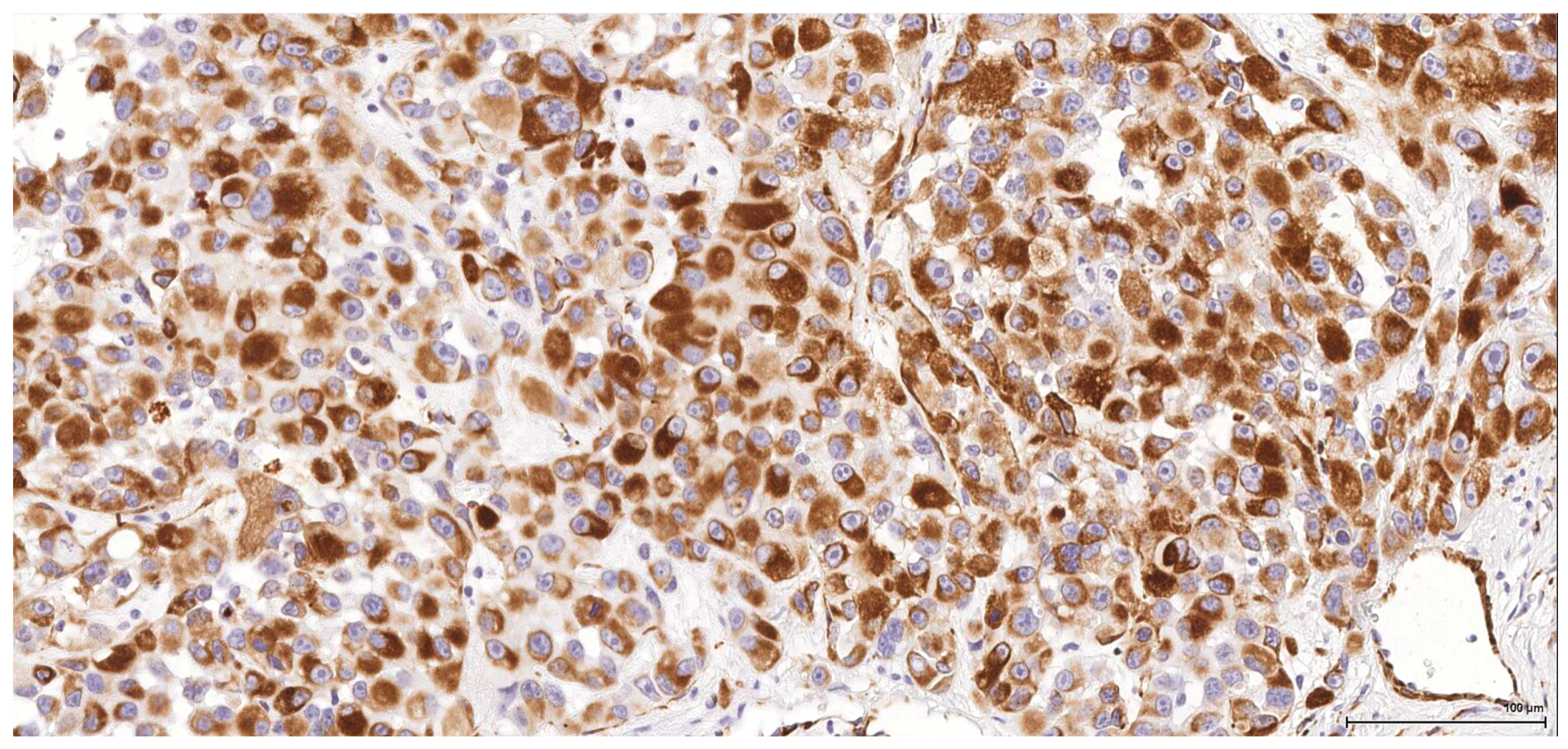
Figure 5. CD10: Male (case 2): intense and diffuse reaction.
- Figure 5 (6 MB)
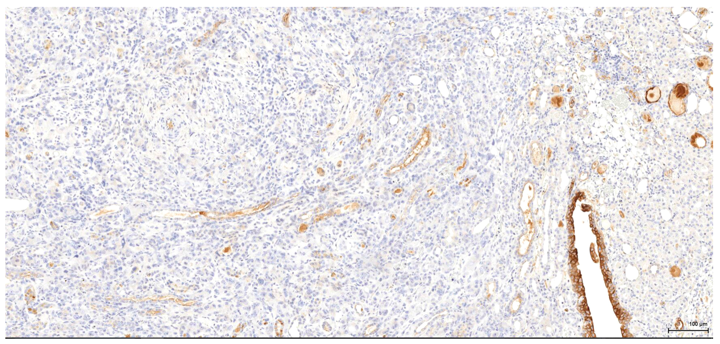
Figure 6. CK7: Male (case 3): tumour cells are negative, whereas residual urothelium is positive.
- Figure 6 (4 MB)
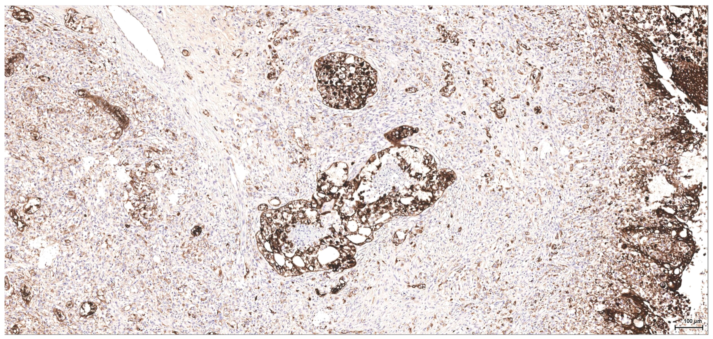
Figure 7. CK MNF116: Female NTP (case 5).
High-grade tumour, a few better differentiated foci with minimal tubular formation.
- Figure 7 (5 MB)
Figure 8. Vimentin: Female NTP (case 5).
Intense and diffuse reaction, including the better-differentiated foci; negative residual urothelium infiltrated by tumour (right).
- Figure 8 (6 MB)
Figure 9. Desmin: Female NTP (case 5).
Rhabdoid features with eccentrically placed nuclei and intense positive reaction.
- Figure 9 (4 MB)
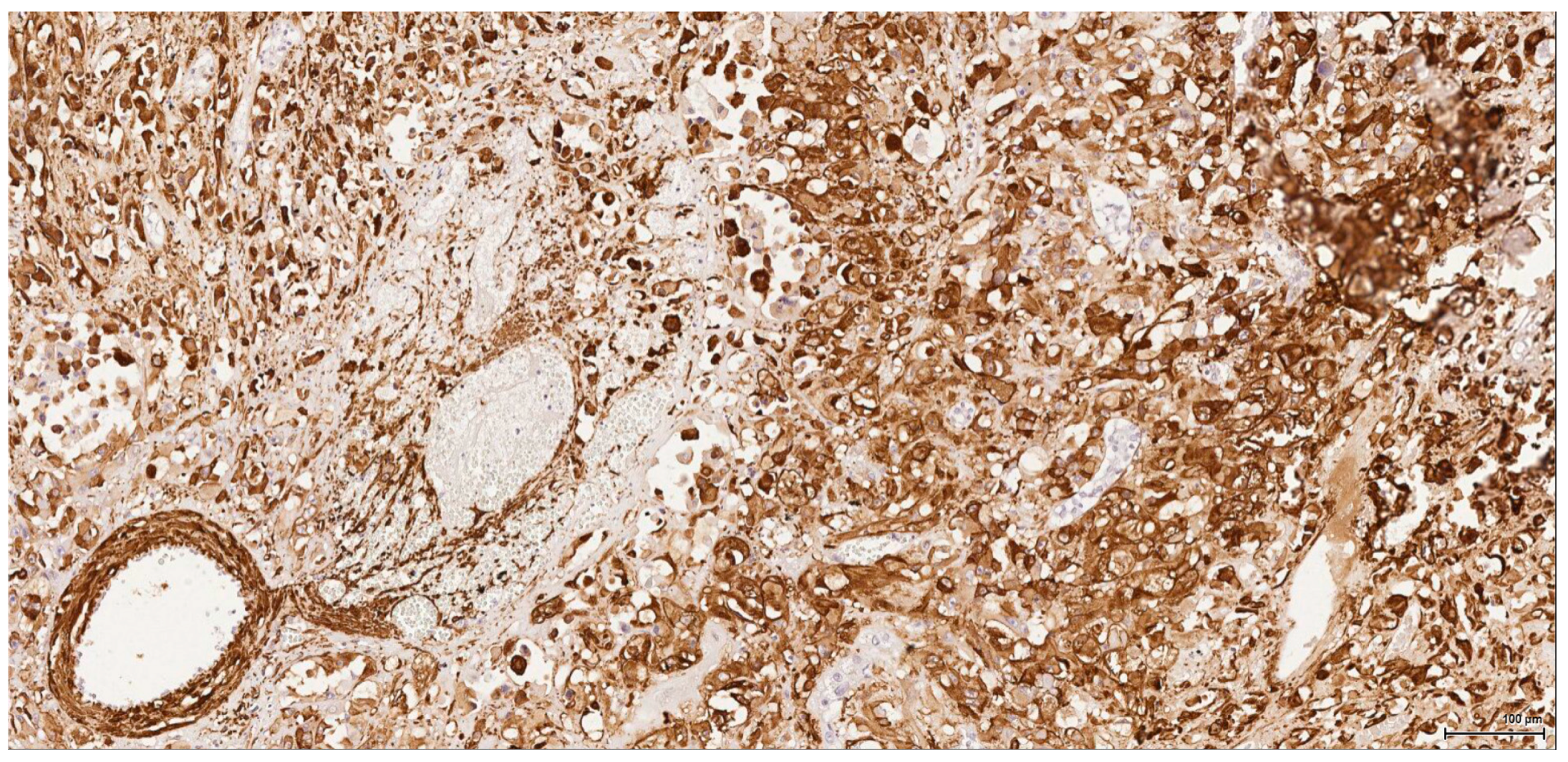
Figure 10. Actin: Male (case 6): intense and diffuse reaction, entrapped negative tubuli.
- Figure 10 (5 MB)
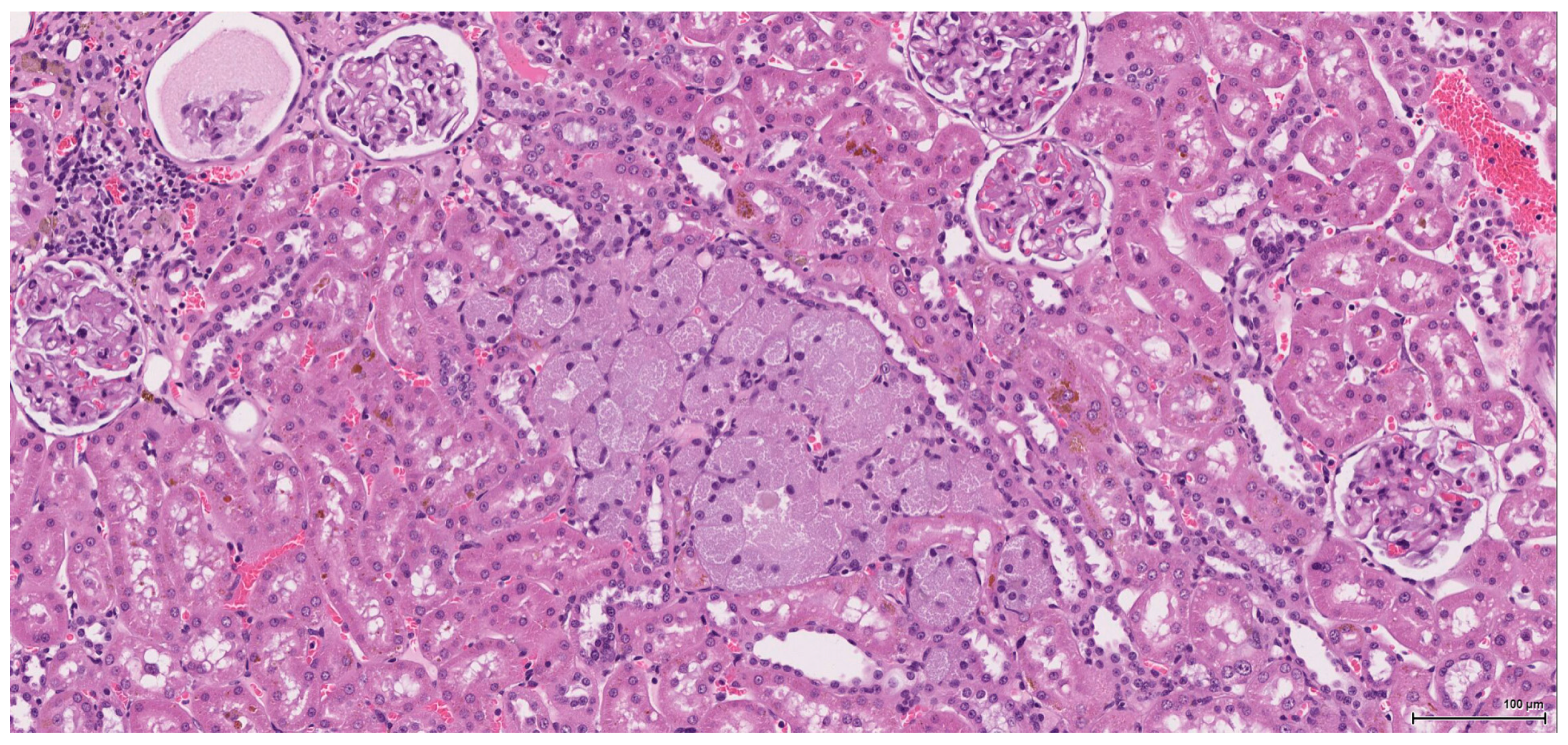
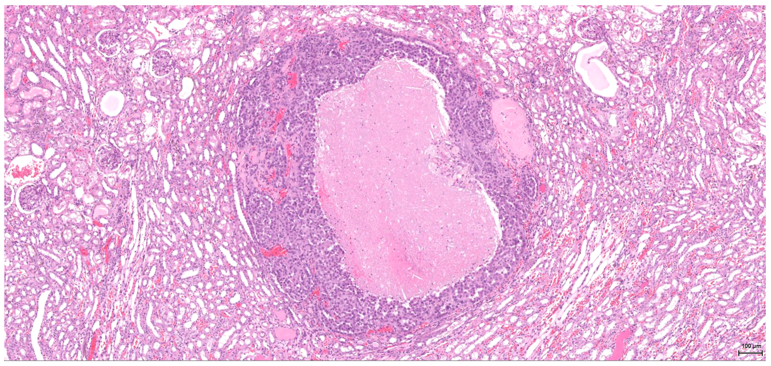
Figure 11. H&E: Female NTP non-tumour case.
Oncocytic hyperplasia (centre) with enlarged, finely granular, pale eosinophilic cytoplasm.
- Figure 11 (5 MB)
Figure 12. H&E: Female NTP tumour case: small, well-differentiated, low-grade tumour.
- Figure 12 (5 MB)
Tables
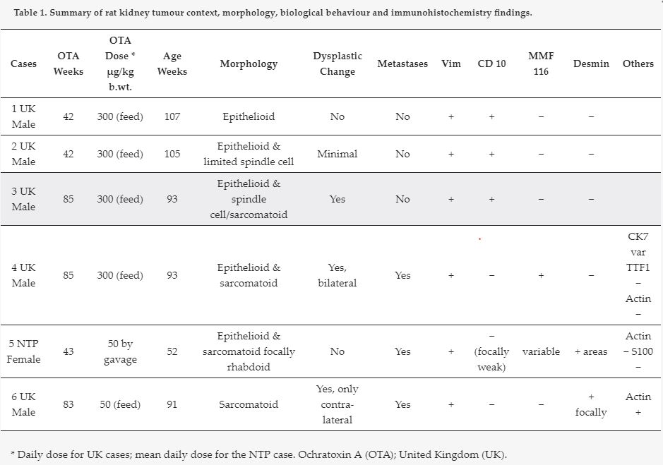
Table 1. Summary of rat kidney tumour context, morphology, biological behaviour and IHC findings.
- Table 1 (78 KB)