

Systems Biology for Organotypic Cell Cultures
Grego S, Dougherty ER, Alexander FJ, Auerbach SS, Berridge BR, Bittner ML, Casey W, Cooley PC, Dash A, Ferguson SS, Fennell TR, Hawkins BT, Hickey AJ, Kleensang A, Liebman MNJ, Martin F, Maull EA, Paragas J, Qiao GG, Ramaiahgari S, Sumner SJ, Yoon M.
ALTEX (2017)
DOI: https://doi.org/10.14573/altex.1608221
PMID: 27846345
Publication
Abstract
Translating in vitro biological data into actionable information related to human health holds the potential to improve disease treatment and risk assessment of chemical exposures. While genomics has identified regulatory pathways at the cellular level, translation to the organism level requires a multiscale approach accounting for intra-cellular regulation, inter-cellular interaction, and tissue/organ-level effects. Tissue-level effects can now be probed in vitro thanks to recently developed systems of three-dimensional (3D), multicellular, "organotypic" cell cultures, which mimic functional responses of living tissue. However, there remains a knowledge gap regarding interactions across different biological scales, complicating accurate prediction of health outcomes from molecular/genomic data and tissue responses. Systems biology aims at mathematical modeling of complex, non-linear biological systems. We propose to apply a systems biology approach to achieve a computational representation of tissue-level physiological responses by integrating empirical data derived from organotypic culture systems with computational models of intracellular pathways to better predict human responses. Successful implementation of this integrated approach will provide a powerful tool for faster, more accurate and cost-effective screening of potential toxicants and therapeutics.
On September 11, 2015, an interdisciplinary group of scientists, engineers, and clinicians gathered for a workshop in Research Triangle Park, North Carolina, to discuss this ambitious goal. Participants represented laboratory-based and computational modeling approaches to pharmacology and toxicology, as well as the pharmaceutical industry, government, non-profits, and academia. Discussions focused on identifying critical system perturbations to model, the computational tools required, and the experimental approaches best suited to generating key data.
Figures
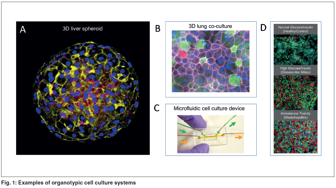
Figure 1. Examples of organotypic cell culture systems.
(A) HepaRG liver cell spheroid stained for actin cytoskeleton and nuclei (red = actin, blue = nuclei).
(B) Primary airway epithelial cells
grown at air-liquid interface (pink = actin, red = mucin, green = cilia, blue = nuclei) in a 3D co-culture with microvascular cells (not shown).
(C) Micromolded microfluidic device for cell culture on a nanoporous membrane.
(D) The HemoShear technology uses cone and
flow viscometer principles to apply hepatic microcirculation flow and transport parameters over 2 weeks. Hepatocytes in this system can
be cultured at physiological insulin concentrations (1000fold lower than in static cultures) resulting in retention of insulin sensitivity
and responses. Hepatocytes develop steatotic changes (Nile Red staining for lipid) under high glucose/high insulin milieus, allowing
modeling of disease-like states, and exhibit drug-induced toxicity phenotypes like steatohepatitis when treated with therapeutic
concentrations of amiodarone. (Panel A courtesy of S. Ferguson, NIEHS/NIH; Panels B-C courtesy of S. Grego, RTI International;
Panel D courtesy of A. Dash, HemoShear)
- Figure 1 (720 KB)
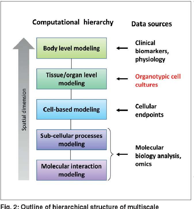
Figure 2. Outline of hierarchical structure of multiscale biological modeling.
Computational models exist for each of the level, having an integrated model linking the "spatial dimensions" is the real challenge.
- Figure 2 (158 KB)
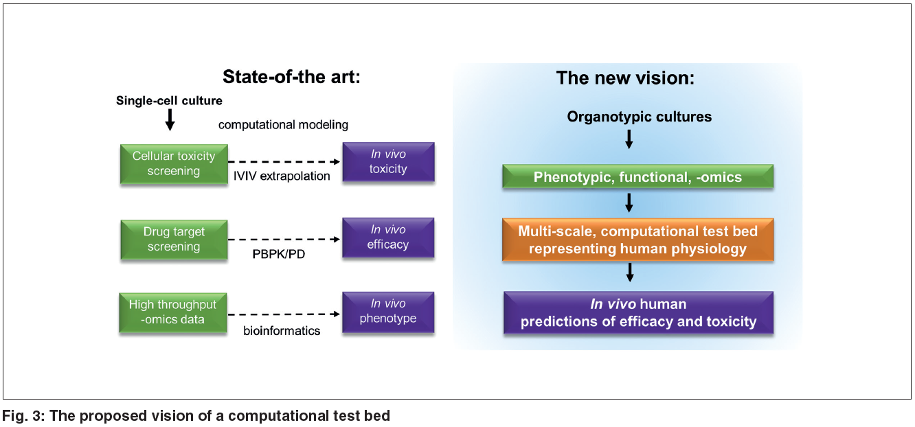
Figure 3. The proposed vision of a computational test bed.
The proposed vision of a computational test bed leverages tissue-level data from organotypic cultures as well as intracellular data to replicate human physiological response across scales and achieve more accurate predictions.
- Figure 3 (277 KB)
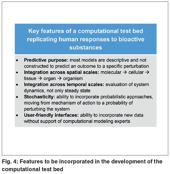
Figure 4. Features to be incorporated in the development of the computational test bed.
- Figure 4 (147 KB)
Figure 5. Disciplines and approaches represented in the workshop.
- Figure 5 (118 KB)