

Airway Injury in an in vitro Human Epithelium-Fibroblast Model of Diacetyl Vapor Exposure: Diacetyl-Induced Basal/Suprabasal Spongiosis
William M. Gwinn, Gordon P. Flake, Ronald W. Bousquet, Genie J. Taylor, and Daniel L. Morgan.
Inhalation Toxicology (2017)
DOI: https://doi.org/10.1080/08958378.2017.1369604
PMID: 28984536
Publication
Abstract
Inhalation exposure to diacetyl (DA) is associated with obliterative bronchiolitis (OB) in workers and induces OB-like fibrotic airway lesions in rats. The pathogenesis of OB is poorly understood in part due to complex interactions between airway epithelial, mesenchymal and blood-derived inflammatory cells. DA-induced airway toxicity in the absence of recruited-inflammatory/immune cells was characterized using an air-liquid interface (ALI) model consisting of human airway epithelium with (Epi/FT) and without (Epi) a mesenchymal component. ALI cultures were exposed to 25 mM DA-derived vapors (using vapor cups) for 1 h on day 0, 2 and 4. In some experiments, the tissues were exposed to 2,3-hexanedione (Hex) which is structurally-similar, but much less fibrogenic than DA. Lactate dehydrogenase activity and day 6 histopathologic changes associated with epithelial injury, including basal/suprabasal spongiosis, were increased following exposure of Epi/FT tissues to DA but not control or Hex vapors. IL-1a, IL-6, IL-8, sIL-1Ra, TGFa, MCP-3 and TNFa proteins were increased following DA exposure of Epi/FT tissues; only IL-1a, IL-8, sIL-1Ra and TGFa were increased following exposure of Epi tissues. MMP-1, MMP-3 and TIMP-1 proteins were increased following DA exposure of Epi/FT tissues; whereas MMP-2, MMP-7 and TIMP-2 were decreased, and production was largely dependent upon the presence of sub-epithelial stromal matrix/fibroblasts. Hex-induced protein changes were minimal. This in vitro study demonstrated that exposure of human airways to DA vapors induced epithelial injury (with the histopathologic feature of basal/suprabasal spongiosis) and increased release of pro-inflammatory and pro-fibrotic cytokines/chemokines as well as MMPs/TIMPs in the absence of recruited-inflammatory cells.
Figures
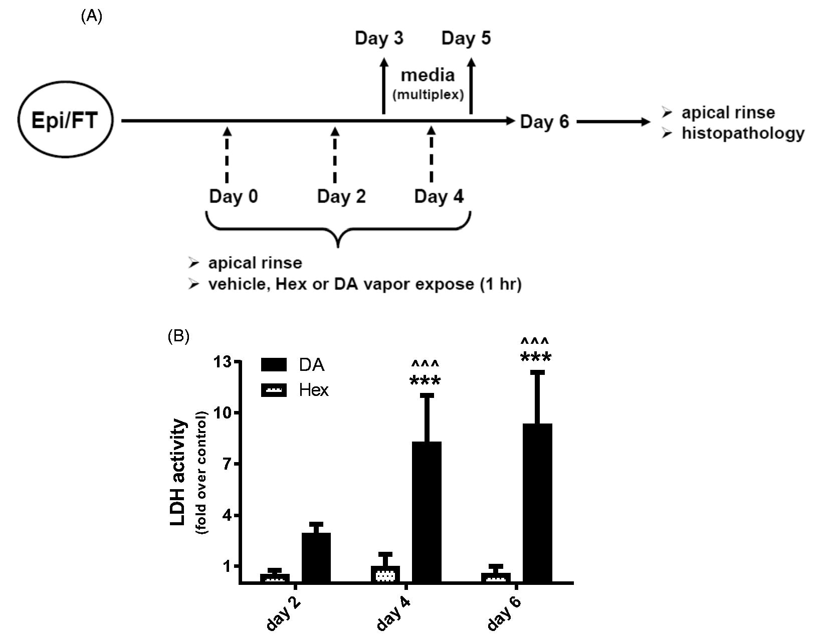
Figure 1. DA vapor exposure induces LDH release.
Epi/FT tissues were exposed to vehicle (PBS), 10 mM Hex- or 25 mM DA-derived vapors (∼1000 ppm) for 1 h on day 0, 2 and 4 as described in Materials and Methods. The experimental design is shown in (A). (B) LDH activity was measured in day 2, 4 and 6 apical rinse supernatants as a marker of cellular injury. Absorbance values from DA or Hex vapor-treated tissues were divided by the mean corresponding value from vehicle-treated tissues and expressed as fold over control. Data is presented as mean ± SD (for graph; n = 6 for vehicle and Hex, n = 12 for DA). The statistics comparing DA versus Hex on each day are shown on graph based on two-way ANOVA with Sidak’s test (^^^p < .001). The statistics comparing DA exposure on day 4 and 6 versus day 2 are also shown based on two-way ANOVA with Tukey’s test (***p < .001).
- Figure 1 (116 KB)
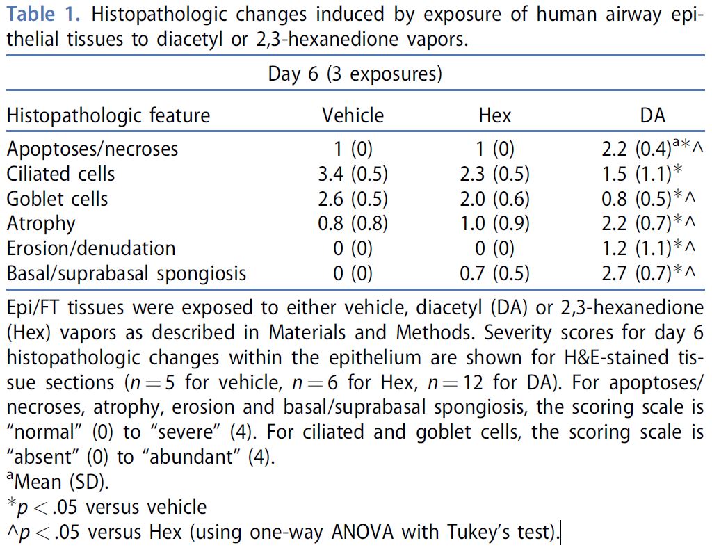
Figure 2. DA vapor exposure induces airway epithelial injury with associated histopathologic changes
DA vapor exposure induces airway epithelial injury with associated histopathologic changes including basal/suprabasal spongiosis. Epi/FT tissues were exposed to vehicle, Hex or DA vapors as described in Materials and Methods. The tissues were fixed in NBF and then processed for histologic evaluation. Representative regions from two separate ALI tissues (same donor) are shown for each type of exposure (A,B) Vehicle control. The epithelial cells are well-differentiated and uniform with many ciliated cells (short arrows) and interspersed goblet cells (long arrows). The deep border (arrowheads) of the epithelium is smooth and the basal cell layer is intact. (C,D) DA vapor-exposed. Goblet cells are absent, and cilia are shortened (short arrows) or absent. Individual cell death (white arrows) is common and characterized by the presence of scattered cells with shrunken, pyknotic nuclei and hypereosinophilic cytoplasm (apoptotic bodies or individual cell necrosis). The basal cell layer contains dying or dead cells, and there are spaces between the adjacent basal cells (basal spongiosis) (arrowheads) as well as a space or cleft between the basal cells and the overlying second row of nuclei (suprabasal spongiosis). In D, the scattered pyknotic cells in the underlying stroma are believed to be degenerate basal epithelial cells that have drifted into the loose, watery matrix. (E,F) Hex vapor-exposed. The tissue in E appears relatively normal with retention of goblet cells (long arrow) and ciliated cells (short arrow), as well as an intact basal cell layer (arrowheads). This is representative of the majority of Hex-vapor treated tissues. However, in the tissue shown in F, there are reduced numbers of goblet cells and ciliated cells (short arrow), occasional apoptotic bodies (white arrow), and apparent degenerative changes within the basal cell layer (arrowheads) resembling spongiosis and clefting. These changes were noted focally in only one of the Hex vapor-treated tissues and were less severe than in the DA vapor-treated tissues. H&E, 40× original objective magnification, all photos.
- Figure 2 (2 MB)
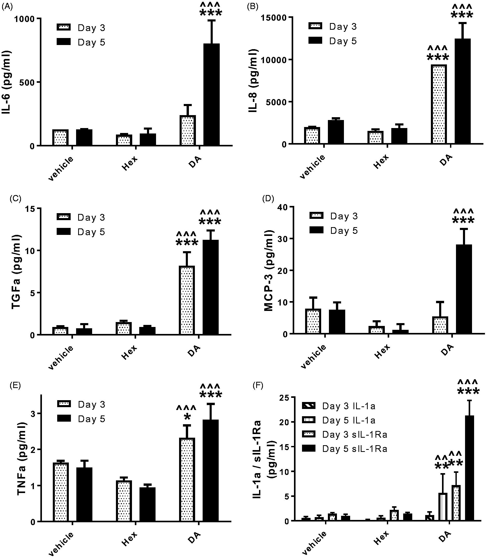
Figure 3. DA vapor exposure induces changes in cytokine/chemokine production.
Epi/FT tissues were exposed to vehicle, Hex or DA vapors as described in Materials and Methods. Multiplex analysis was used to measure a panel of cytokines/chemokines in day 3 and 5 culture media samples. (A) IL-6, (B) IL-8, (C) TGFa, (D) MCP-3, (E) TNFa, (F) IL-1a and sIL-1Ra are shown. Data is presented as mean ± SD (for graphs; n = 3 for vehicle and Hex, n = 6 for DA). Only the statistics comparing all three treatment groups on each day are shown on graphs based on two-way ANOVA with Tukey’s test (*p < .05, **p < .01, ***p < .001 versus vehicle; ^^p < .01, ^^^p < .001 versus Hex).
- Figure 3 (365 KB)
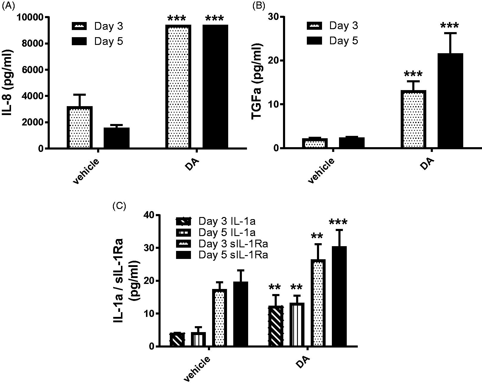
Figure 4. DA vapor-induced production of IL-8, TGFa, IL-1a and sIL-1Ra.
DA vapor-induced production of IL-8, TGFa, IL-1a and sIL-1Ra does not require the presence of mesenchymal component. Donor (TBE-20)-matched tissues without matrix/fibroblasts (Epi) were exposed to vehicle or DA vapors as described in Materials and Methods. Multiplex analysis was used to measure a panel of cytokines/chemokines in day 3 and 5 culture media samples. (A) IL-8, (B) TGFa, (C) IL-1a and sIL-1Ra are shown. Data is presented as mean ± SD (for graphs; n = 4 for vehicle and DA). Only the statistics comparing DA versus vehicle on each day are shown on graphs based on two-way ANOVA with Sidak’s test (**p < .01, ***p < .001).
- Figure 4 (211 KB)
Figure 5. DA vapor exposure induces changes in MMP/TIMP production.
DA vapor exposure induces changes in MMP/TIMP production. Epi/FT tissues were exposed to vehicle, Hex or DA vapors as described in Materials and Methods. Multiplex analysis was used to measure a panel of MMPs and TIMPs in day 5 culture media samples. (A) MMP-1, (B) MMP-3, (C) MMP-2, (D) MMP-7, (E) TIMP-1 and (F) TIMP-2 are shown. Data is presented as mean ± SD (for graphs; n = 3 for vehicle and Hex, n = 6 for DA). The statistics comparing all three treatment groups are shown on graphs based on one-way ANOVA with Tukey’s test (*p < .05, **p < .01, ***p < .001 versus vehicle; ^p < .05, ^^p < .01, ^^^p < .001 versus Hex).
- Figure 5 (265 KB)
Tables
Table 1. Histopathologic changes induced by exposure of human airway epithelial tissues.
Histopathologic changes induced by exposure of human airway epithelial tissues to diacetyl or 2,3-hexanedione vapors.
- Table 1 (183 KB)
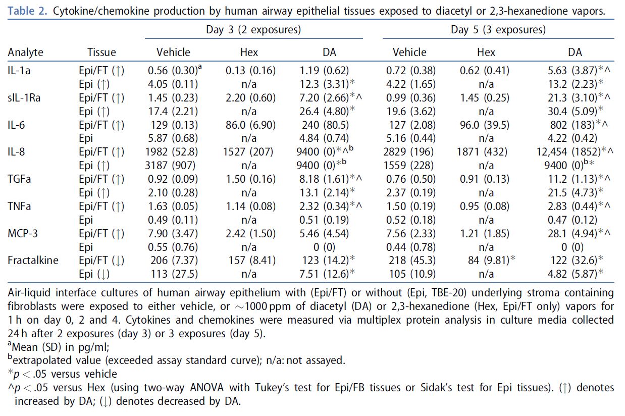
Table 2. Cytokine/chemokine production by human airway epithelial tissues.
Cytokine/chemokine production by human airway epithelial tissues exposed to diacetyl or 2,3-hexanedione vapors.
- Table 2 (248 KB)
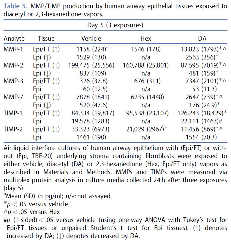
Table 3. MMP/TIMP production by human airway epithelial tissues.
MMP/TIMP production by human airway epithelial tissues exposed to diacetyl or 2,3-hexanedione vapors.
- Table 3 (168 KB)