

Molecular Effects of 1-Naphthyl-Methylcarbamate and Solar Radiation Exposures on Human Melanocytes
Bianca Ferrucio, Manoela Tiago, Richard D. Fannin, Liwen Liu, Kevin Gerrish, Silvya Stuchi Maria-Engler, Richard S. Paules, and Silvia Berlanga de Moraes Barros.
Toxicology in Vitro (2017)
DOI: https://doi.org/10.1016/j.tiv.2016.11.005
PMID: 27829164
Publication
Abstract
Carbaryl (1-naphthyl-methylcarbamate), a broad-spectrum insecticide, has recently been associated with the development of cutaneous melanoma in an epidemiological cohort study with U.S. farm workers also exposed to ultraviolet radiation, the main etiologic factor for skin carcinogenesis. We hypothesized that carbaryl exposure may increase deleterious effects of UV solar radiation on skin melanocytes. This study aimed to characterize human melanocytes after individual or combined exposure to carbaryl (100μM) and solar radiation (375mJ/cm2). In a microarray analysis, carbaryl, but not solar radiation, induced an oxidative stress response, evidenced by the upregulation of antioxidant genes, such as Hemeoxygenase-1 (HMOX1), and downregulation of Microphtalmia-associated Transcription Factor (MITF), the main regulator of melanocytic activity; results were confirmed by qRT-PCR. Carbaryl and solar radiation induced a gene response suggestive of DNA damage and cell cycle alteration. The expression of CDKN1A, BRCA1/2 and MDM2 genes was notably more intense in the combined treatment group, in a synergistic manner. Flow cytometry assays demonstrated S-phase cell cycle arrest, reduced apoptosis levels and faster induction of cyclobutane pyrimidine dimers (CPD) lesions in carbaryl treated groups. Our data suggests that carbaryl is genotoxic to human melanocytes, especially when associated with solar radiation.
Figures
Figure 1. Relative gene expression validation by qRT-PCR.
Relative gene expression validation by qRT-PCR – comparison between microarray and qRT-PCR data. Data generated in experimental triplicate, analyzed by ANOVA followed by Dunnett's test to evaluate differences between treated groups and the control group, and ANOVA followed by Tukey's test to verify which groups differ from one another (*p < 0.05, **p < 0.001). Results are expressed as fold change (mean ± standard deviation) relative to DMSO treatment (vehicle control). Groups identified by the same letter do not differ significantly. Circled asterisks indicate a synergistic response evaluated by Student t-test for the non-orthogonal contrast (p < 0.001), between Carbaryl and UV radiation treatments. UV – treated with DMSO 0.07% and 375 mJ/cm2 solar radiation; Carb UV – treated with carbaryl 100 μM and 375 mJ/cm2 solar radiation; Carb – treated with carbaryl 100 μM; DMSO – treated with DMSO 0.07%.
- Figure 1 (372 KB)
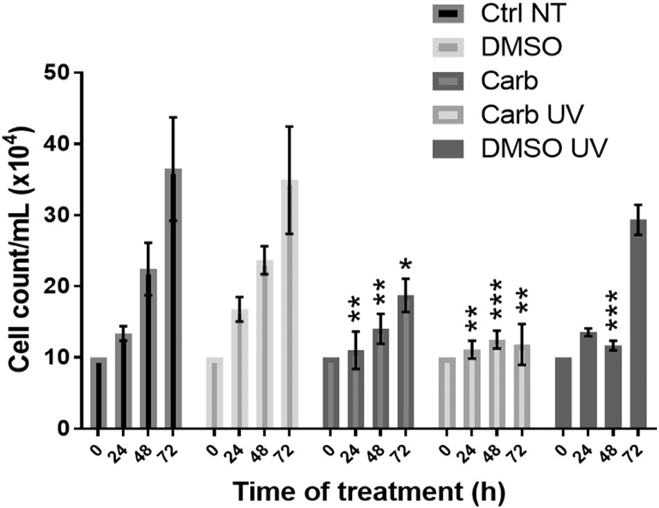
Figure 2. Cell growth evaluation - cell counting after 24, 48 and 72 h of treatment.
Data was generated in triplicate, analyzed by Data generated in experimental triplicate, analyzed by ANOVA followed by Dunnett's test to evaluate differences between treated groups and the control group (*p < 0.05; **p < 0.005; ***p < 0.0005). DMSO UV – treated with DMSO 0.07% and irradiated with solar radiation 375 mJ/cm2; Carb UV – treated with carbaryl 100 μM and irradiated with solar radiation 375 mJ/cm2; Carb – treated with carbaryl 100 μM; DMSO – treated with DMSO 0.07%; and Ctrl – non-treated group.
- Figure 2 (99 KB)
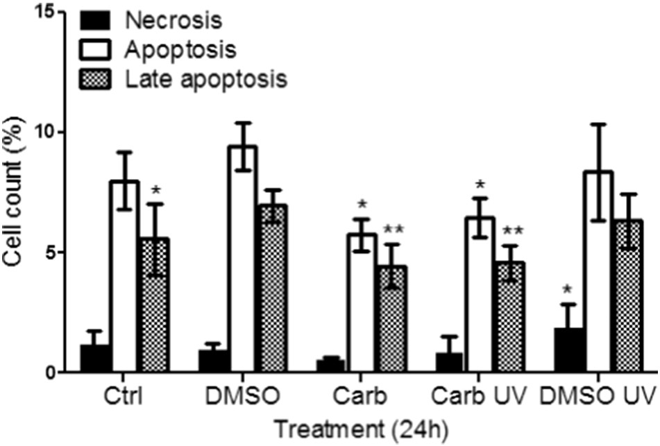
Figure 3. Evaluation of cell death mechanisms by flow cytometry analysis – human melanocytes.
Evaluation of cell death mechanisms by flow cytometry analysis – human melanocytes treated for 24 h. Data was generated in triplicate, analyzed by ANOVA followed by Tukey's test, and compared to the DMSO control group (*p < 0.05, **p < 0.001). DMSO UV – treated with DMSO 0.07% and irradiated with solar radiation 375 mJ/cm2; Carb UV – treated with carbaryl 100 μM and irradiated with solar radiation 375 mJ/cm2; Carb – treated with carbaryl 100 μM; DMSO – treated with DMSO 0.07%; and Ctrl – non-treated group.
- Figure 3 (88 KB)
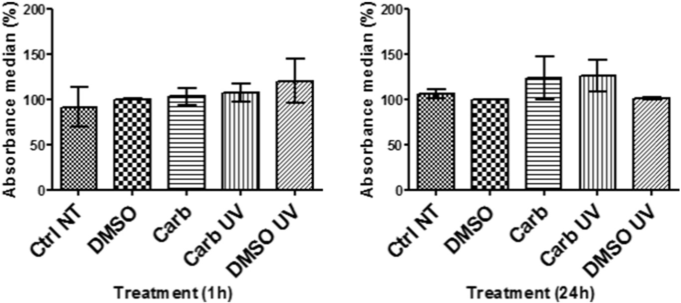
Figure 4. Quantification of cyclobutane pyrimidine dimers (CPDs).
Quantification of cyclobutane pyrimidine dimers (CPDs): fluorescence absorbance median analyzed by flow cytometry after 1 and 6 h of treatment. Data was generated in triplicate, analyzed by ANOVA followed by the Tukey's test, and compared to the DMSO control group (*p < 0.05, **p < 0.005).
DMSO UV – treated with DMSO 0.07% and irradiated with solar radiation 375 mJ/cm2; Carb UV – treated with carbaryl 100 μM and irradiated with solar radiation 375 mJ/cm2; Carb – treated with carbaryl 100 μM; DMSO – treated with DMSO 0.07%; and Ctrl – non-treated group.
- Figure 4 (125 KB)
Figure 5. Quantification of 8-oxo-dG: fluorescence median absorbance analyzed by flow cytometry.
Quantification of 8-oxo-dG: fluorescence median absorbance analyzed by flow cytometry after 1 and 24 h of treatment. Data was generated in triplicate, analyzed by ANOVA followed by the Tukey's test, and compared to the DMSO control group.
DMSO UV – treated with DMSO 0.07% and irradiated with solar radiation 375 mJ/cm2; Carb UV – treated with carbaryl 100 μM and irradiated with solar radiation 375 mJ/cm2; Carb – treated with carbaryl 100 μM; DMSO – treated with DMSO 0.07%; and Ctrl – non-treated group.
- Figure 5 (126 KB)
Tables
Table 1. Significantly altered canonical pathways and the related altered genes.
Significantly altered canonical pathways and the related altered genes in the different treatment groups.
- Table 1 (179 KB)
Table 2. Evaluation of cell cycle phase at different time-points.
- Table 2 (160 KB)
Supplemental Materials
Supplementary Data
- Figure S1-S2 (164 KB)