

Binding of Bisphenol A, Bisphenol AF, and Bisphenol S on the Androgen Receptor: Coregulator Recruitment and Stimulation of Potential Interaction Sites
Lalith Perera, Yin Li, Laurel A. Coons, Rene Houtman, Rinie van Beuningen, Bonnie Goodwin, Scott S. Auerbach, and Christina T. Teng.
Toxicology in Vitro (2017)
DOI: https://doi.org/10.1016/j.tiv.2017.07.020
PMID: 28751236
Publication
Abstract
Bisphenol A (BPA), bisphenol AF (BPAF), and bisphenol S (BPS) are well known endocrine disruptors. Previous in vitro studies showed that these compounds antagonize androgen receptor (AR) transcriptional activity; however, the mechanisms of action are unclear. In the present study, we investigated interactions of coregulator peptides with BPA, BPAF, or BPS at the AR complexes using Micro Array for Real-time Coregulator Nuclear Receptor Interaction (MARCoNI) assays and assessed the binding of these compounds on AR by molecular dynamics (MD) simulations. The set of coregulator peptides that are recruited by BPA-bound AR, either positively/or negatively, are different from those recruited by the agonist R1881-bound AR. Therefore, the data indicates that BPA shows no similarities to R1881 and suggests that it may recruit other coregulators to the AR complex. BPAF-bound AR recruits about 70-80% of the same coregulator peptides as BPA-bound AR. Meanwhile, BPS-bound AR interacts with only few peptides compared to BPA or BPAF-bound AR. MD results show that multiple binding sites with varying binding affinities are available on AR for BPA, BPAF, and BPS, indicating the availability of modified binding surfaces on AR for coregulator interactions. These findings help explain some of the distinct AR-related toxicities observed with bisphenol chemicals and raise concern for the use of substitutes for BPA in commercial products.
Figures
Figure 1. The profiles of coregulator peptides interacting with AR using a MARCoNI assay.
The profiles of coregulator peptides interacting with AR (bound to R1881, CPA, BPA, BPAF, and BPS) using a MARCoNI assay.
(A) Working model of the MARCoNI assay.
(B) The heatmap of the peptide screening (Red color bar shows positive interactions and blue color bar shows negative interactions)
(C) Modulation Index (MI) in response to R1881, CPA, BPA, BPAF, and BPS binding to the AR.
(For interpretation of the references to color in this figure legend, the reader is referred to the web version of this article.)
- Figure 1 (2 MB)
Figure 2. Comparison of the coregulatory-derived binding peptides.
Comparison of the coregulatory-derived binding peptides when ligands R1881, CPA, BPA, BPAF, and BPS are bound to the AR.
(A) Positive interactions. (B) Negative interactions. (C) Overlapping positive interactions. (D) Overlapping negative interactions.
- Figure 2 (1 MB)
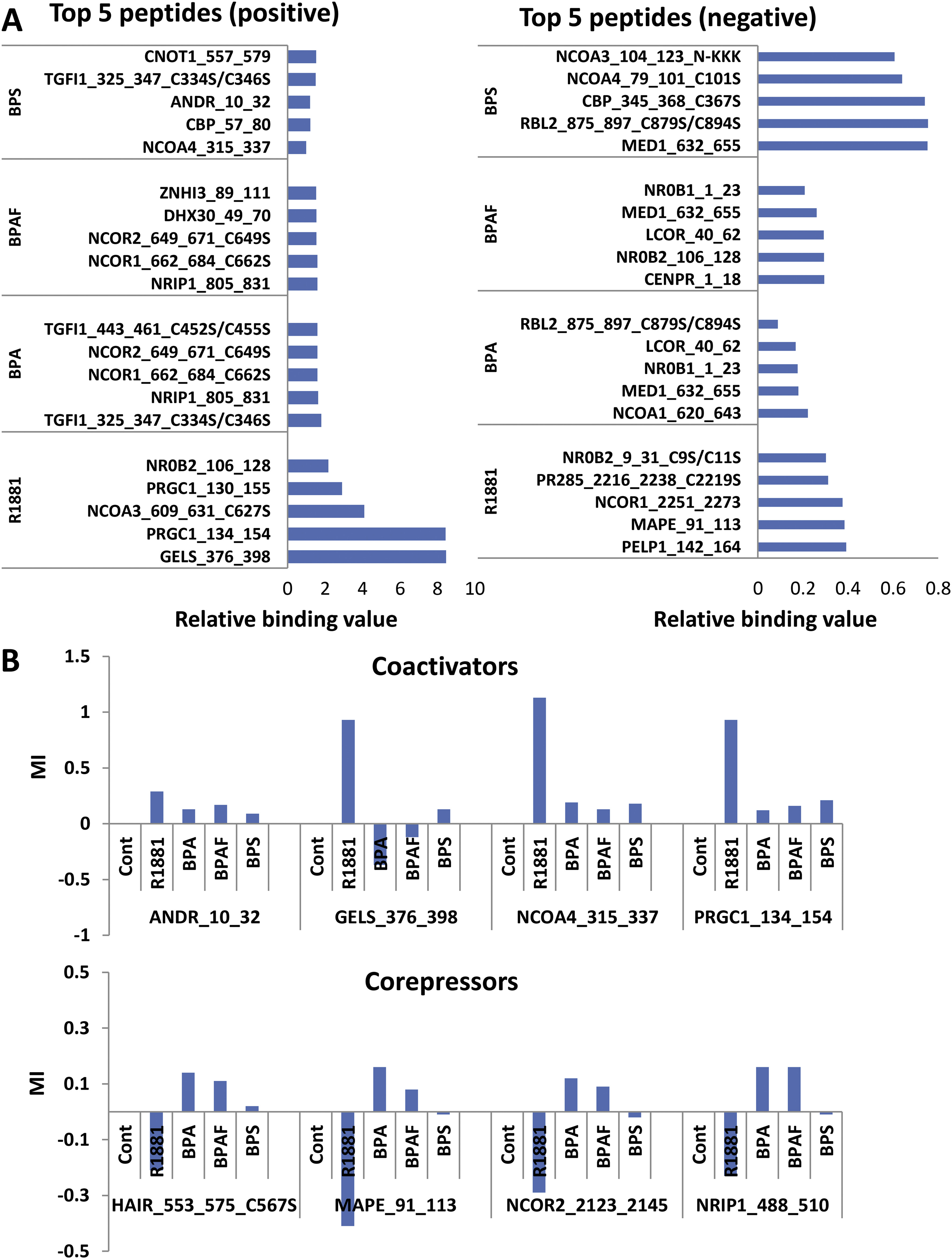
Figure 3. Primary binding peptides and coregulators.
(A) Top 5 positive and negative peptides. (B) Top 5 positive and negative coactivators and corepressors.
- Figure 3 (1 MB)
Figure 4. Chemical structures of the ligands (agonists and BPA analogues) used in the MD simulations.
Various binding sites of BPA analogues on AR tested in the present MD study are shown in the bottom-left. For comparison, the SRC binding site of DHT bound AR from the X-ray crystal structure (pdb ID 3L3X) is given in the bottom-right. The three helices, H3, H5 (behind AF2), and H12 that create the binding surface of coregulators are also shown.
- Figure 4 (675 KB)
Figure 5. Representative solution structures from the latter part of the MD simulations and the corresponding electrostatic potential surface (EPS).
Representative solution structures (left) from the latter part of the MD simulations of ligand-bound AR where the ligand is at the natural LBS and the corresponding electrostatic potential surface (EPS) (right) of ligand-bound AR. Locations of ligands are labelled. Orientation of LBD-AR in all cases is preserved to facilitate the comparison.
- Figure 5 (2 MB)
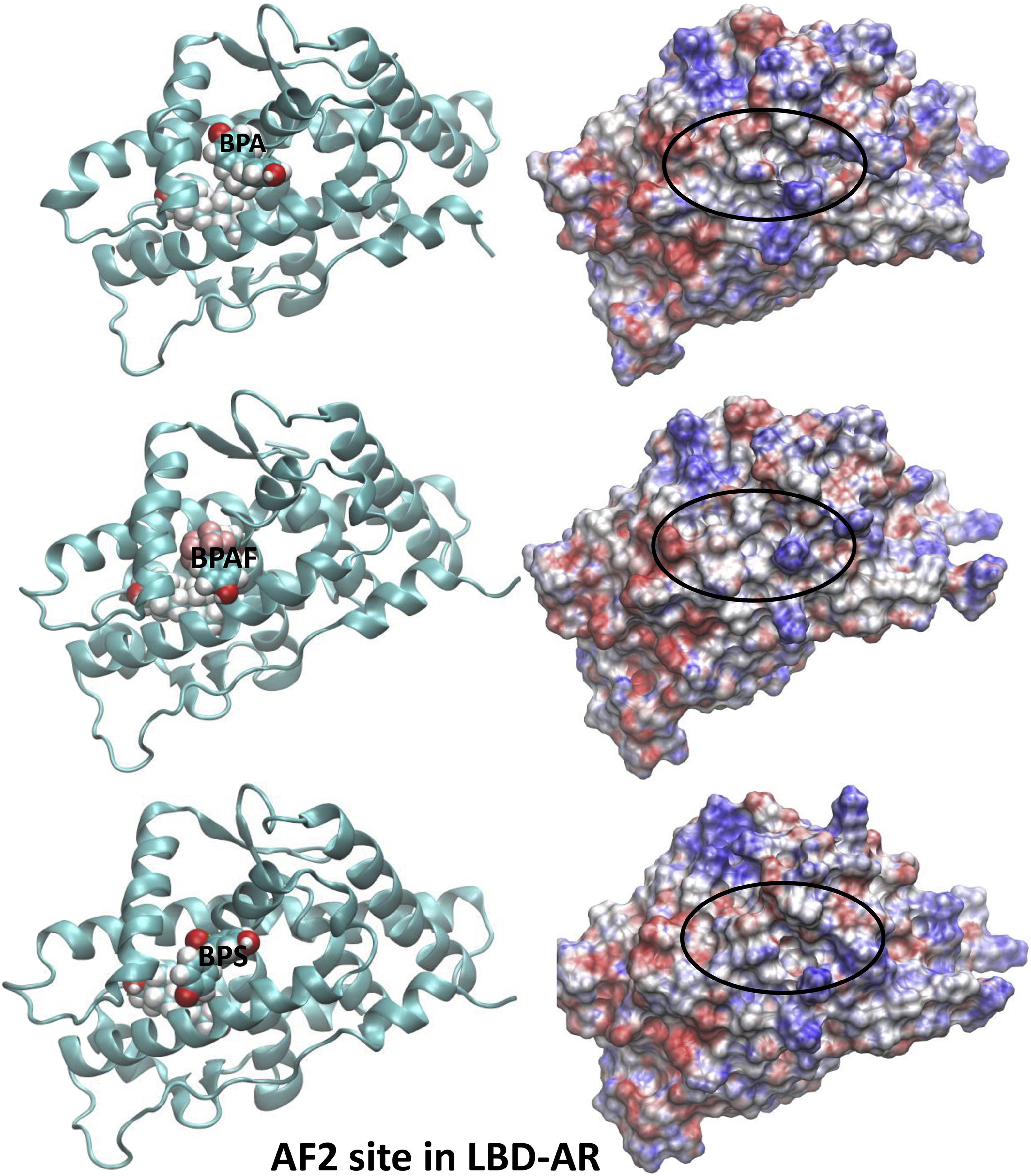
Figure 6. Representative solution structures and the corresponding EPS (at the AF2 site of LBD-AR).
Representative solution structures (left) of BPA analogues-bound AR from MD simulations and the corresponding EPS (right) of ligand-AR complexes where the BPA analogues are at the AF2 site of LBD-AR. In all cases, a DHT molecule is placed at LBS.
- Figure 6 (2 MB)
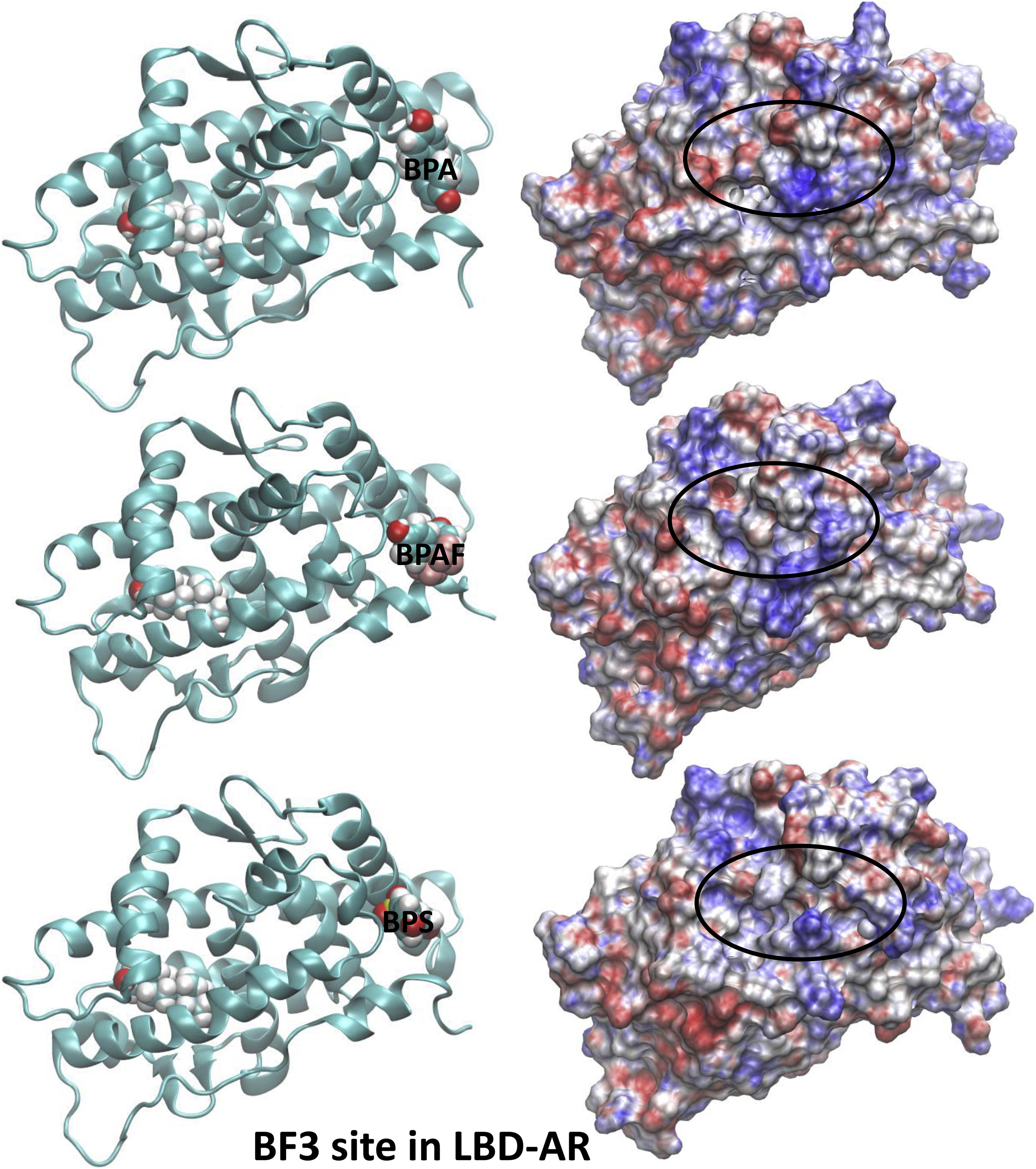
Figure 7. Representative solution structures and the corresponding EPS (at the BF3 site of LBD-AR).
Representative solution structures (left) of BPA analogues-bound AR from MD simulations and the corresponding EPS (right) of ligand-AR complexes where the BPA analogues are at the BF3 site of LBD-AR. In all cases, a DHT molecule is placed at LBS.
- Figure 7 (2 MB)
Tables
Table 1
Table 1a. 27 peptides that positively interact with R1881.
Table 1b. 31 peptides that positively interact with CPA.
Table 1c. 45 peptides that positively interact with BPA.
Table 1d. 44 peptides that positively interact with BPAF.
Table 1e. 4 peptides that positively interact with BPS.
- Table 1 (64 KB)
Table 2
Table 2a. 51 peptides that negatively interact with R1881.
Table 2b. 18 peptides that negatively interact with CPA.
Table 2c. 34 peptides that negatively interact with BPA.
Table 2d. 31 peptides that negatively interact with BPAF.
Table 2e. 12 peptides that negatively interact with BPS.
- Table 2 (134 KB)
Table 3. Binding enthalpies (kcal/mol) of the ligands on AR calculated using MD simulations.
- Table 3 (80 KB)
Supplemental Materials
Supplementary Data
- Supplementary Materials (2 MB)