

Black Cohosh Extracts and Powders Induce Micronuclei, A Biomarker of Genetic Damage, in Human Cells
Stephanie L. Smith‐Roe, Carol D. Swartz, Kim G. Shepard, Steven M. Bryce, Stephen D. Dertinger, Suramya Waidyanatha, Grace E. Kissling, Scott S. Auerbach, Kristine L. Witt, and E. Zeiger.
Environmental and Molecular Mutagenesis (2018)
DOI: https://doi.org/10.1002/em.22182
PMID: 29668046
DOI: https://doi.org/10.22427/NTP-DATA-002-01731-0030-0000-7
Publication
Abstract
Black cohosh extract (BCE) is a widely used dietary supplement marketed to women to alleviate symptoms of gynecological ailments, yet its toxicity has not been well characterized. The National Toxicology Program (NTP) previously reported significant increases in micronucleated erythrocytes in peripheral blood of female Wistar Han rats and B6C3F1/N mice administered 15–1,000 mg BCE/kg/day by gavage for 90 days. These animals also developed a dose‐dependent nonregenerative macrocytic anemia characterized by clinical changes consistent with megaloblastic anemia. Both micronuclei (MN) and megaloblastic anemia can arise from disruption of the folate metabolism pathway. The NTP used in vitro approaches to investigate whether the NTP's test lot of BCE, BCEs from various suppliers, and root powders from BC and other cohosh species, were genotoxic in general, and to gain insight into the mechanism of action of BCE genotoxicity. Samples were tested in human TK6 lymphoblastoid cells using the In Vitro MicroFlow® MN assay. The NTP BCE and a BC extract reference material (XRM) were tested in the MultiFlow® DNA Damage assay, which assesses biomarkers of DNA damage, cell division, and cytotoxicity. The NTP BCE and several additional BCEs were tested in bacterial mutagenicity assays. All samples induced MN when cells were grown in physiological levels of folic acid. The NTP BCE and BC XRM produced activity patterns consistent with an aneugenic mode of action. The NTP BCE and five additional BCEs were negative in bacterial mutagenicity tests. These findings show that black cohosh preparations induce chromosomal damage and may pose a safety concern.
Figures
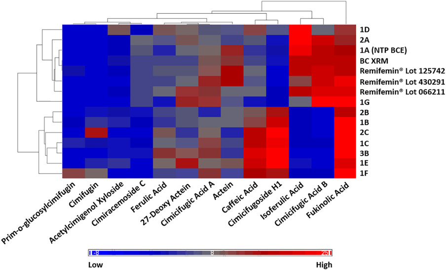
Figure 1. Heatmap analysis of cohosh samples using hierarchical clustering.
Each cell depicts the % area under the peak for the 13 constituents used to characterize the cohosh samples.
- Figure 1 (87 KB)
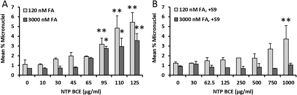
Figure 2. TK6 cells were incubated with the NTP BCE for 24 hr in cell culture medium.
TK6 cells were incubated with the NTP BCE for 24 hr in cell culture medium containing either 120 or 3,000 nM FA in the absence (A) or presence (B) of S9. Each concentration was evaluated using triplicate wells. Error bars represent one standard error above the mean. Significant trend tests: 120 nM FA, P = 0.004; 3,000 nM FA, P < 0.001; 120 nM FA with S9, P < 0.001. For pairwise comparisons, * P < 0.025, ** P < 0.01.
- Figure 2 (56 KB)
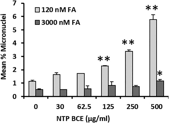
Figure 3. TK6 cells were incubated with the NTP BCE for 4 hr in cell culture medium.
TK6 cells were incubated with the NTP BCE for 4 hr in cell culture medium containing either 120 or 3,000 nM FA. Each concentration was evaluated using duplicate wells. Error bars represent one standard error above the mean. Significant trend test: 120 nM FA, P < 0.001. For pairwise comparisons, * P < 0.025, ** P < 0.01.
- Figure 3 (38 KB)
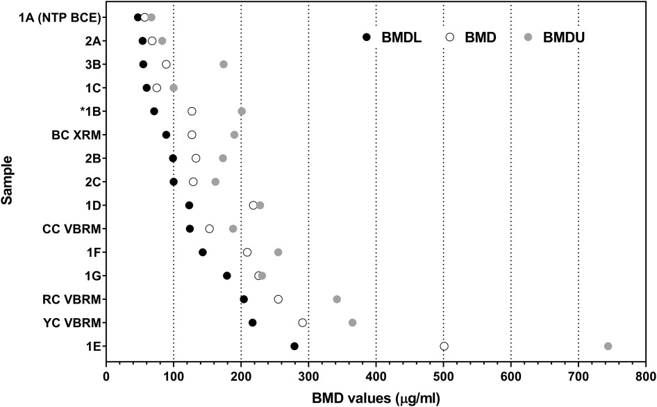
Figure 4. BMD analysis of cohosh sample dose‐response curves ranked in order of lowest to highest BMDL.
*The dose‐response data for BCE sample 1B did not meet the criteria for the goodness of fit parameter when analyzed using BMDExpress2 software.
- Figure 4 (68 KB)
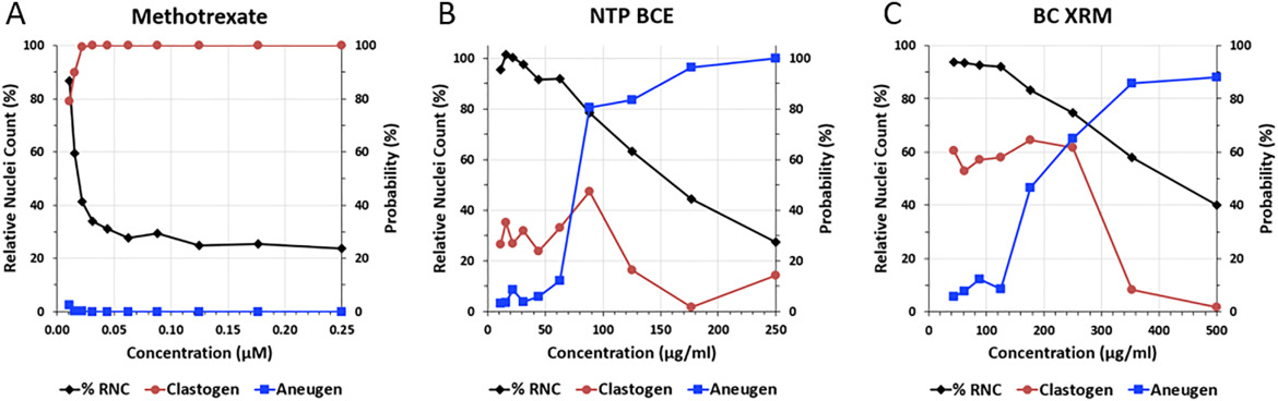
Figure 5. TK6 cell MultiFlow DNA Damage assay predictive algorithm results.
TK6 cell MultiFlow DNA Damage assay predictive algorithm results at each concentration for γH2AX, phospho‐histone H3, p53 translocation, and polyploidy after 4 or 24 hr of exposure, and cytotoxicity after 24 hr of exposure, to methotrexate, a positive control for clastogenicity (A), the NTP BCE (B) and the BC XRM (C). Assays conducted with TK6 cells.
- Figure 5 (101 KB)
Tables
Table 1. Test Article, Supplier, Lot Number, and Identifier.
- Table 1 (129 KB)
Table 2. Micronucleus Assay Results for Black Cohosh Samples (3000 Versus 120 nM Folic Acid in Cell Culture Medium).
- Table 2 (174 KB)
Supplemental Materials
NTP Tables
- G00058D G06 Ames Summary Data (74 KB)
- G00058V G06 Ames Summary Data (68 KB)
- G00058W G06 Ames Summary Data (85 KB)
- G00058X G06 Ames Summary Data (63 KB)
- G00058Y G06 Ames Summary Data (68 KB)
- G00058Z G06 Ames Summary Data (63 KB)
Supporting Information
- Supporting Information (302 KB)