

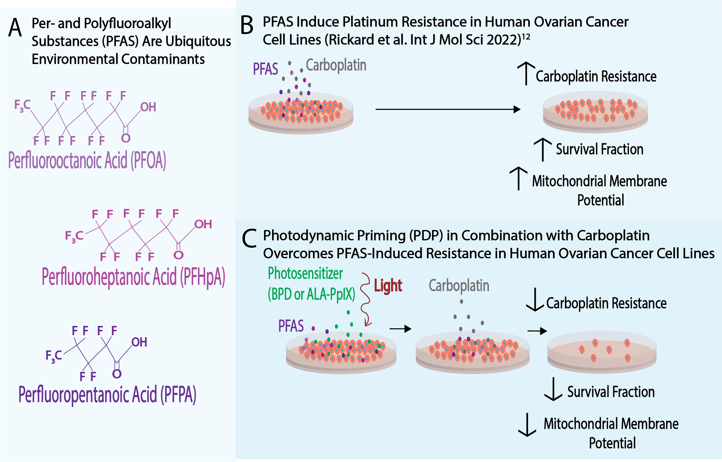
Photodynamic Priming Overcomes Per- and Polyfluoroalkyl Substance (PFAS)-Induced Platinum Resistance in Ovarian Cancer
Brittany P Rickard, Xianming Tan, Suzanne E Fenton, Imran Rizvi
Publication
Publication
Photochemistry & Photobiology DOI: https://doi.org/10.1111/php.13728
DOI:
https://doi.org/10.22427/NTP-DATA-021-00006-0002-0000-1
PMID: 36148678
Abstract
Per- and polyfluoroalkyl substances (PFAS) are widespread environmental contaminants linked to adverse outcomes, including for female reproductive biology and related cancers. We recently reported, for the first time, that PFAS induce platinum resistance in ovarian cancer, potentially through altered mitochondrial function. Platinum resistance is a major barrier in the management of ovarian cancer, necessitating complementary therapeutic approaches. Photodynamic therapy (PDT) is a light-based treatment modality that reverses platinum resistance and synergizes with platinum-based chemotherapy. The present study is the first to demonstrate the ability of photodynamic priming (PDP), a low-dose, sub-cytotoxic variant of PDT, to overcome PFAS-induced platinum resistance. Comparative studies of PDP efficacy using either benzoporphyrin derivative (BPD) or 5-aminolevulinic acid-induced protoporphyrin IX (PpIX) were conducted in two human ovarian cancer cell lines (NIH:OVCAR-3 and Caov-3). BPD and PpIX are clinically approved photosensitizers that preferentially localize to, or are partly synthesized in, mitochondria. PDP overcomes carboplatin resistance in PFAS-exposed ovarian cancer cells, demonstrating the feasibility of this approach to target the deleterious effects of environmental contaminants. Decreased survival fraction in PDP + carboplatin treated cells was accompanied by decreased mitochondrial membrane potential, suggesting that PDP modulates the mitochondrial membrane, reducing membrane potential and re-sensitizing ovarian cancer cells to carboplatin.
Graphical Abstract
- Graphical Abstract (385 KB)
Tables
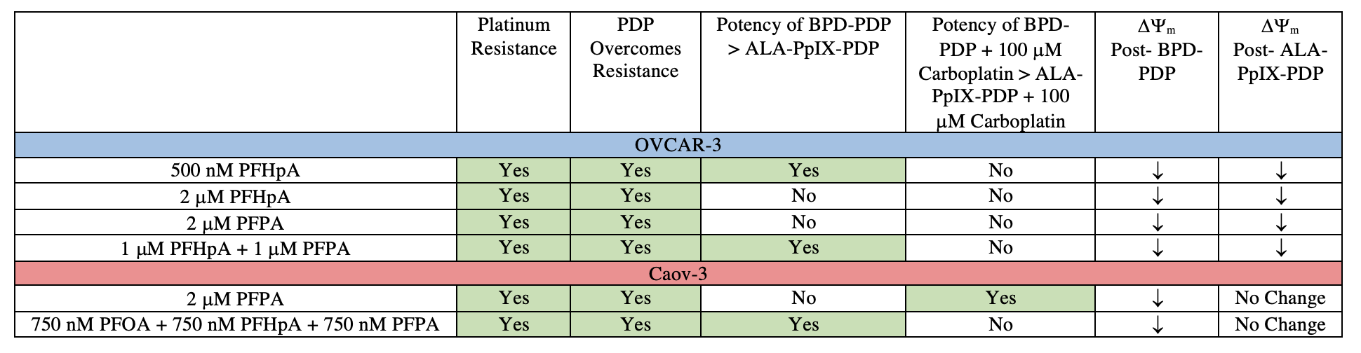
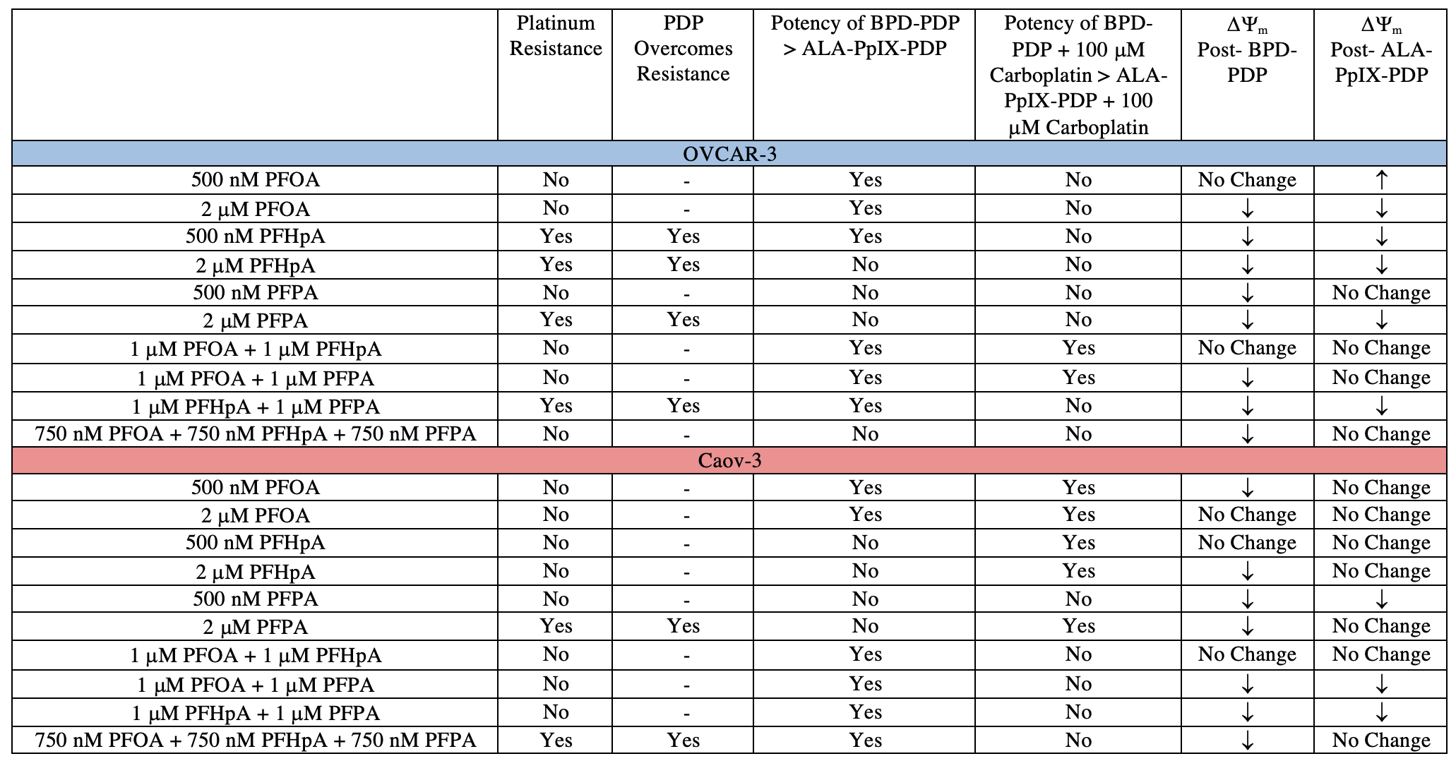
Table 1. Summary of Results
- Table 1 (136 KB)
Figures
All Figures were made from this data file:
- CEBS - PFAS PDT Data (170 KB)
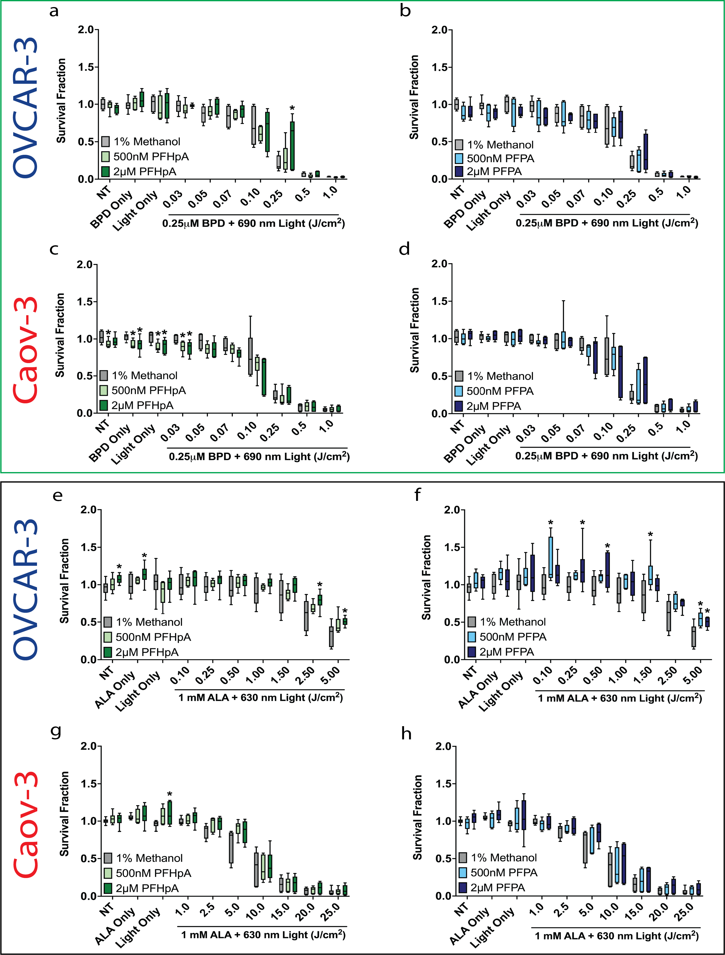
Figure 1. Effect of PFHpA and PFPA on light-dose-dependent responses to BPD-PDT (green box) or ALA-PpIX-PDT (black box) in OVCAR-3 and Caov-3 cells.
- FINAL Figure 1 (747 KB)
- Statistical Analysis 1 (1 MB)
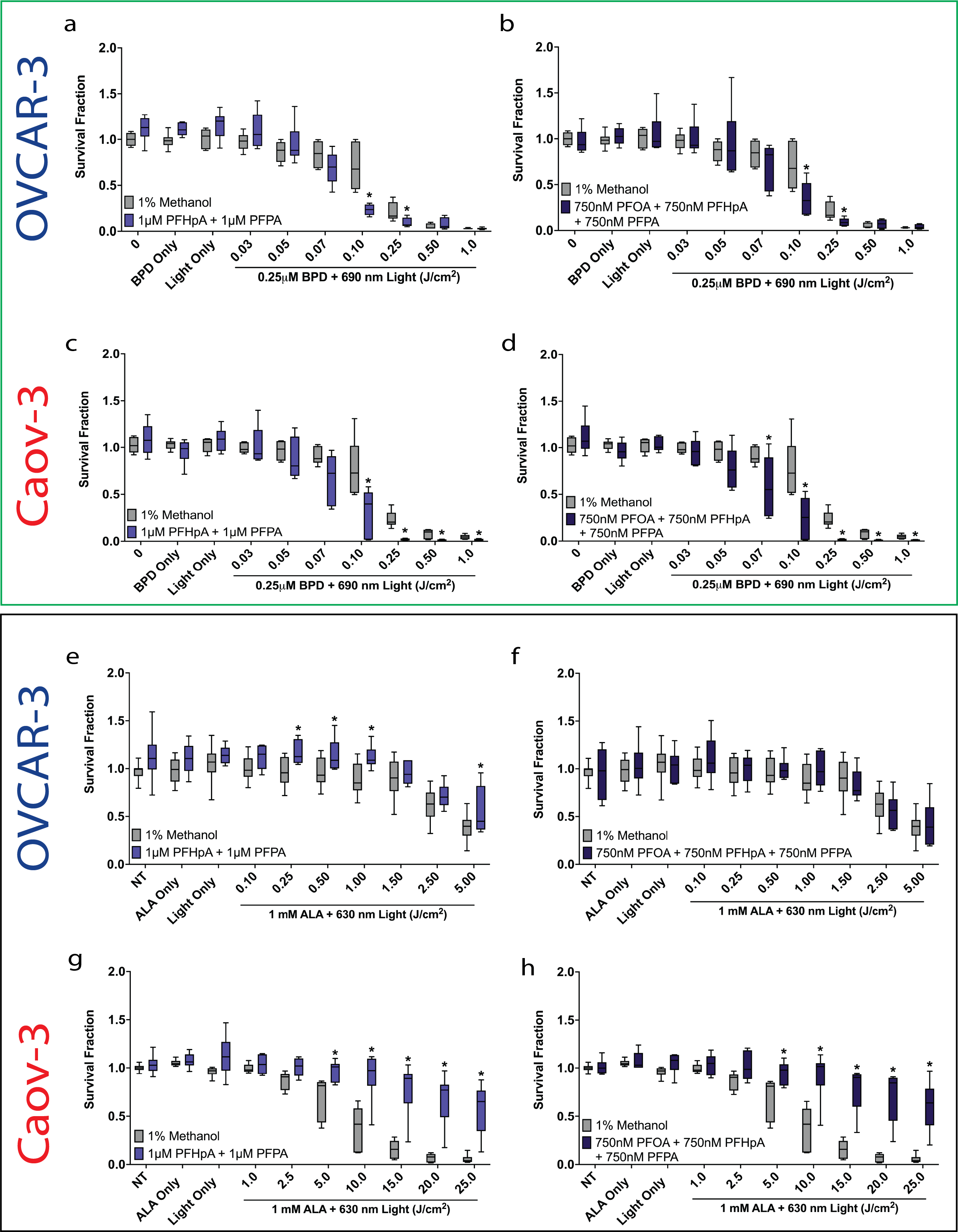
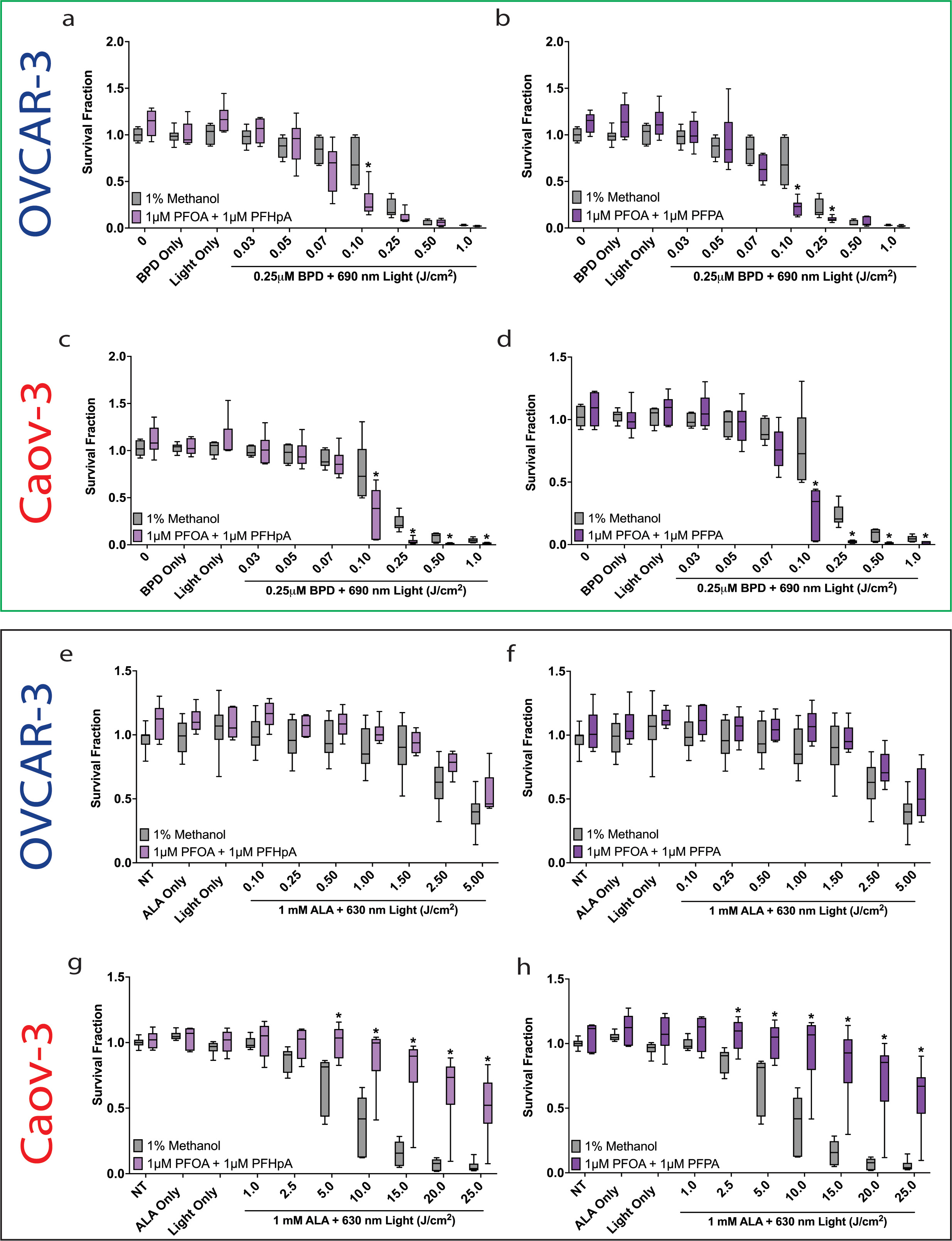
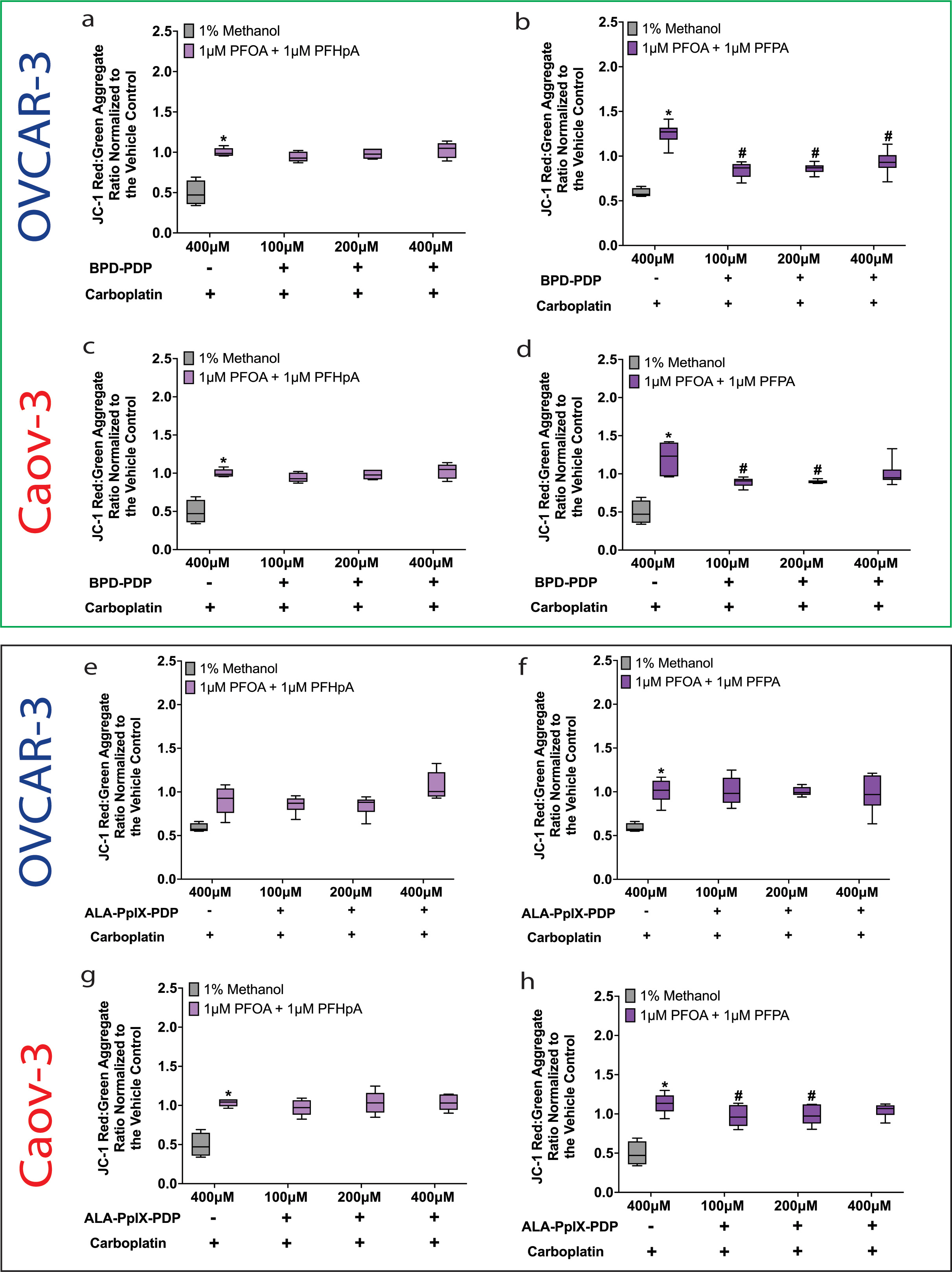
Figure 2. Effect of PFAS mixtures on light-dose-dependent responses to BPD-PDT (green box) or ALA-PpIX-PDT (black box) in OVCAR-3 and Caov-3 cells
- FINAL Figure 2 (640 KB)
- Statistical Analysis 1 (1 MB)
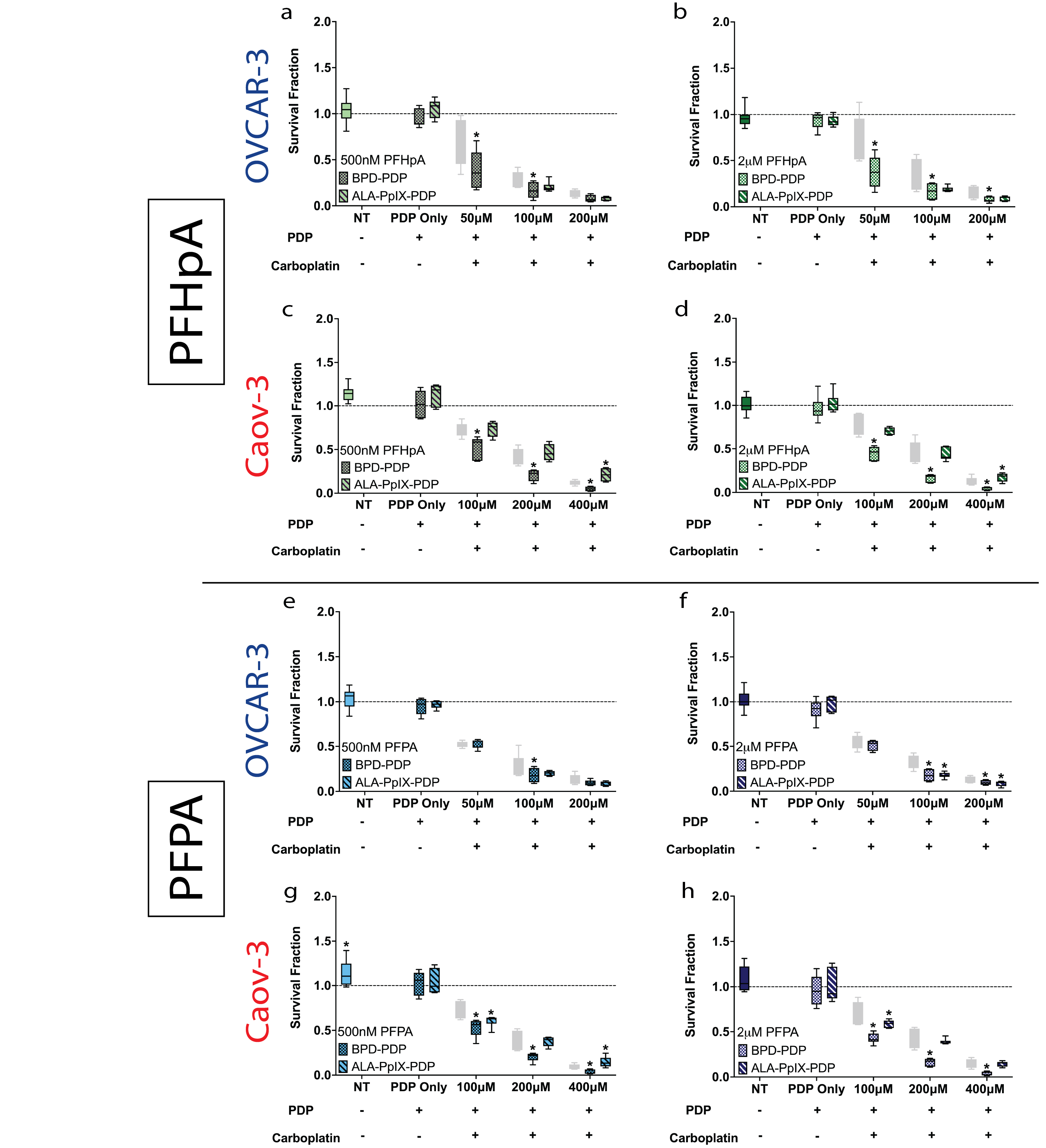
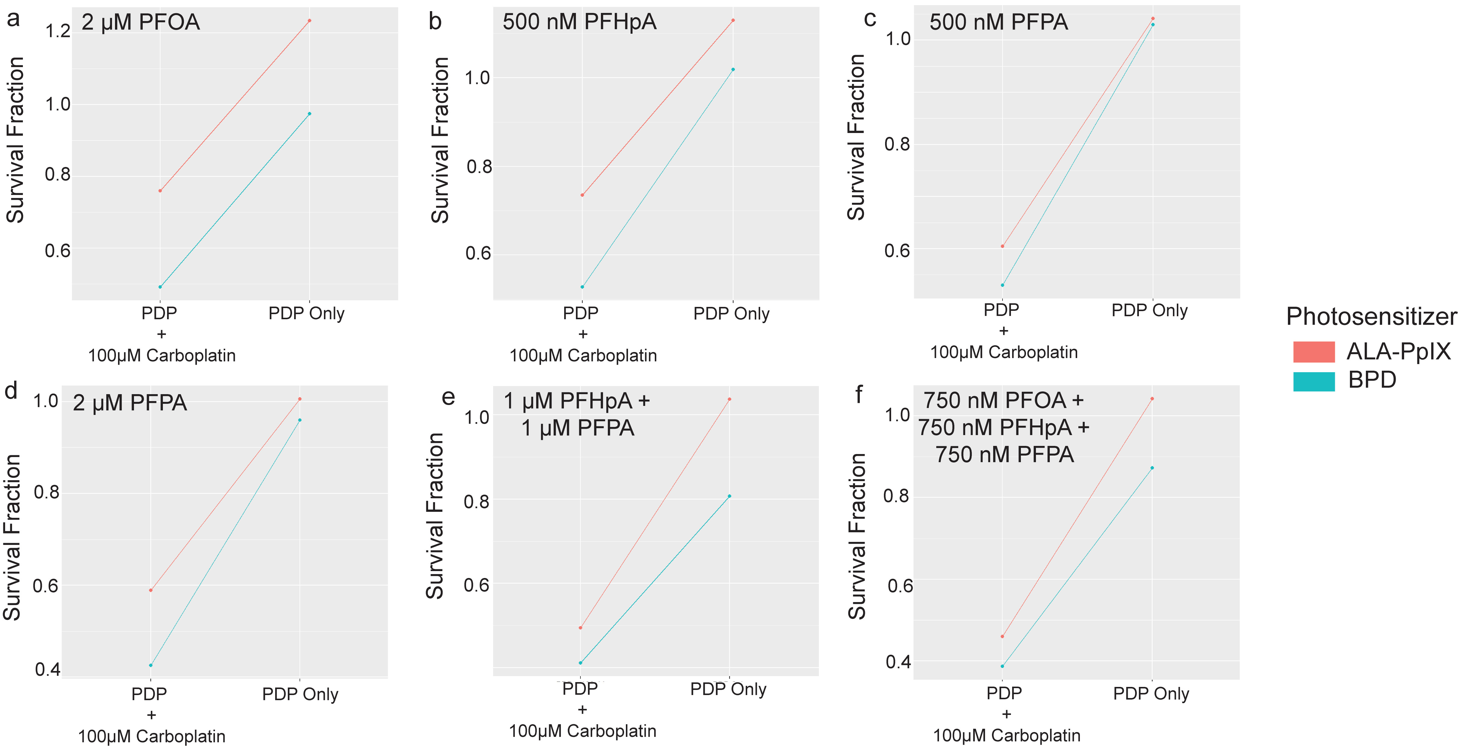
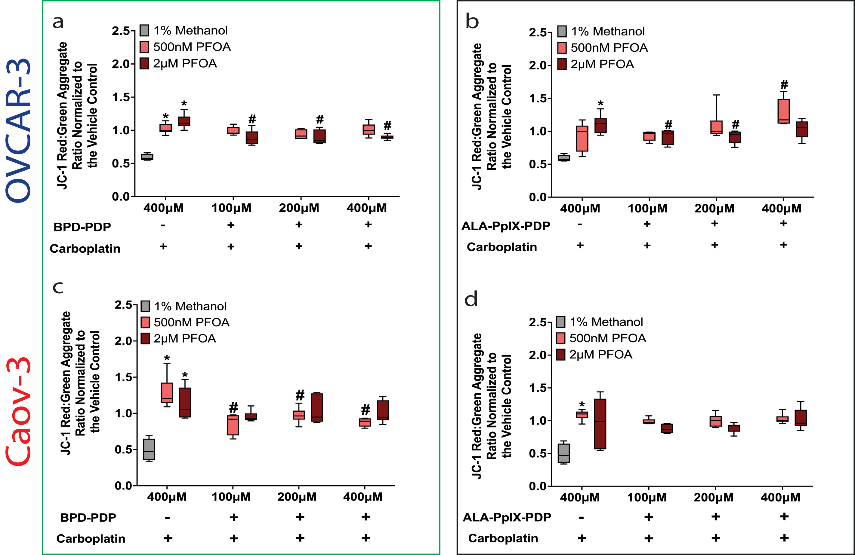
Figure 3. Survival fraction decreased in PFAS-exposed OVCAR-3 and Caov-3 cells post-BPD-PDP or ALA-PpIX-PDP + carboplatin
- FINAL Figure 3 (598 KB)
- Statistical Analysis 2 (1 MB)
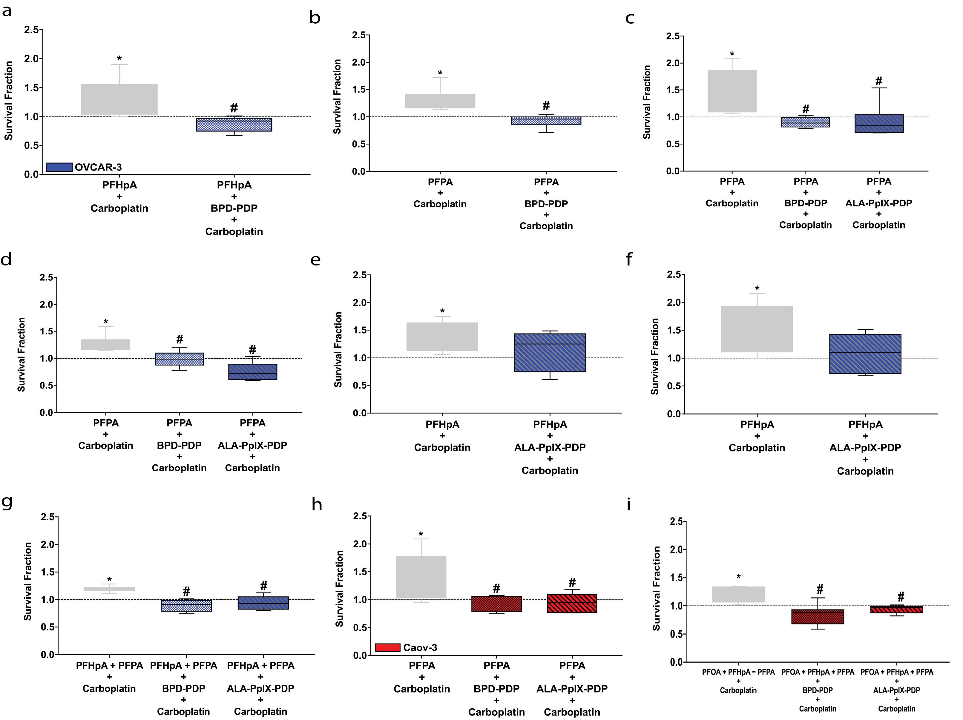
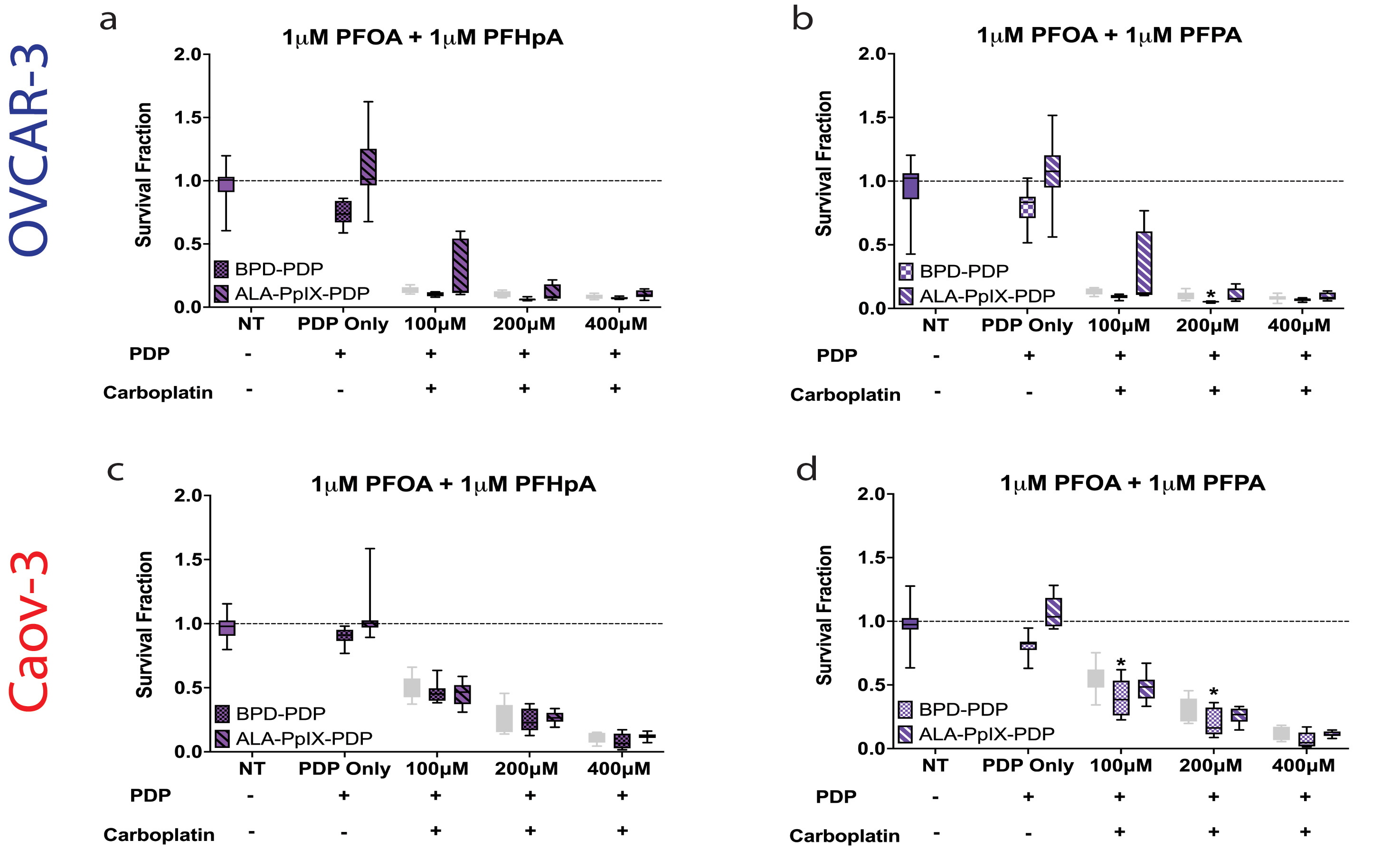
Figure 4. In OVCAR-3 and Caov-3 cells exposed to PFAS mixtures, BPD-PDP was more effective than ALA-PpIX-PDP at reducing survival fraction in combination with carboplatin
- FINAL Figure 4 (357 KB)
- Statistical Analysis 2 (1 MB)
Figure 5. In OVCAR-3 and Caov-3 cell exposure groups where platinum resistance was not observed, photosensitizer efficacy for PDP differed between BPD and ALA-PpIX
- FINAL Figure 5 (224 KB)
- Statistical Analysis 5 (1 MB)
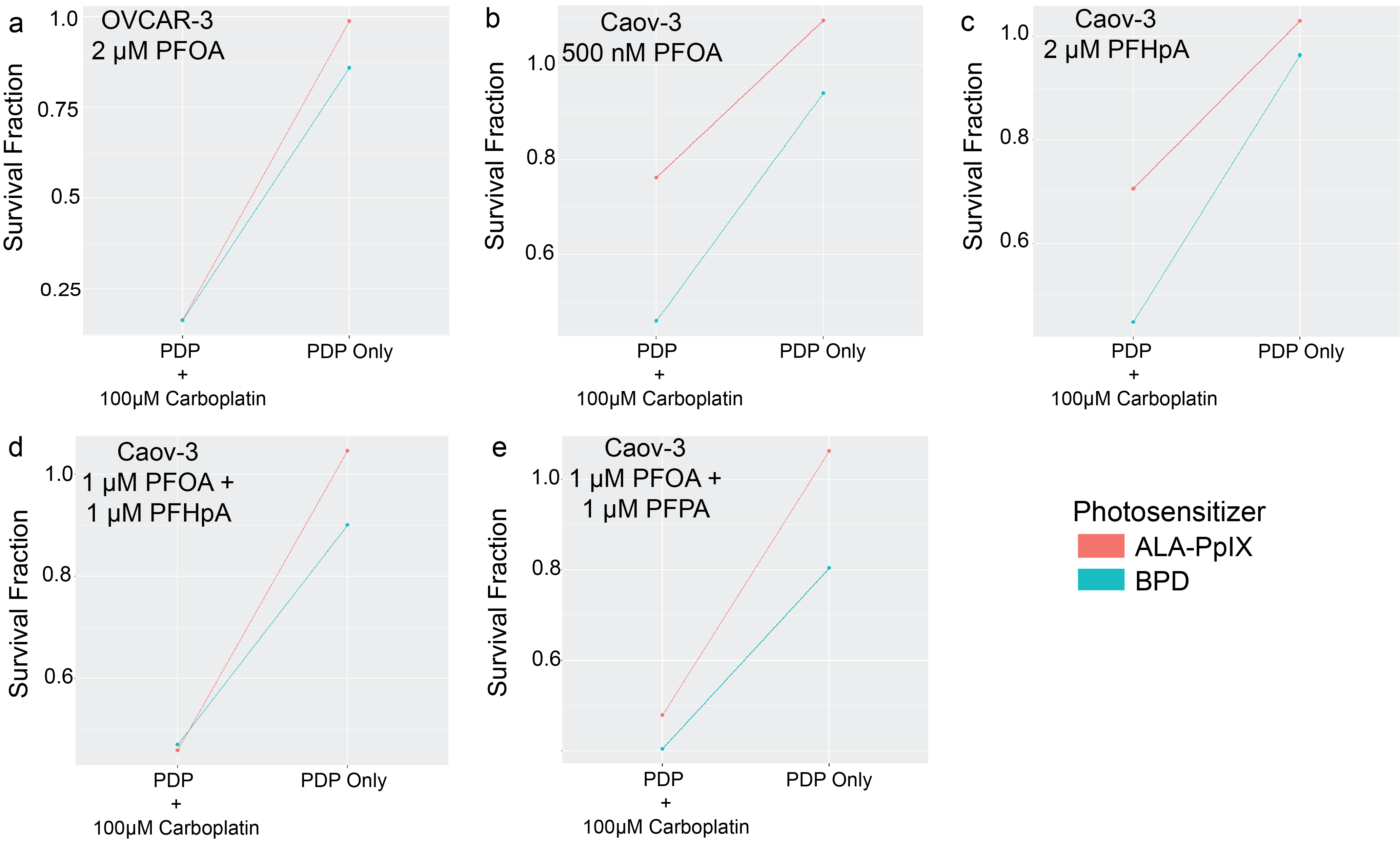
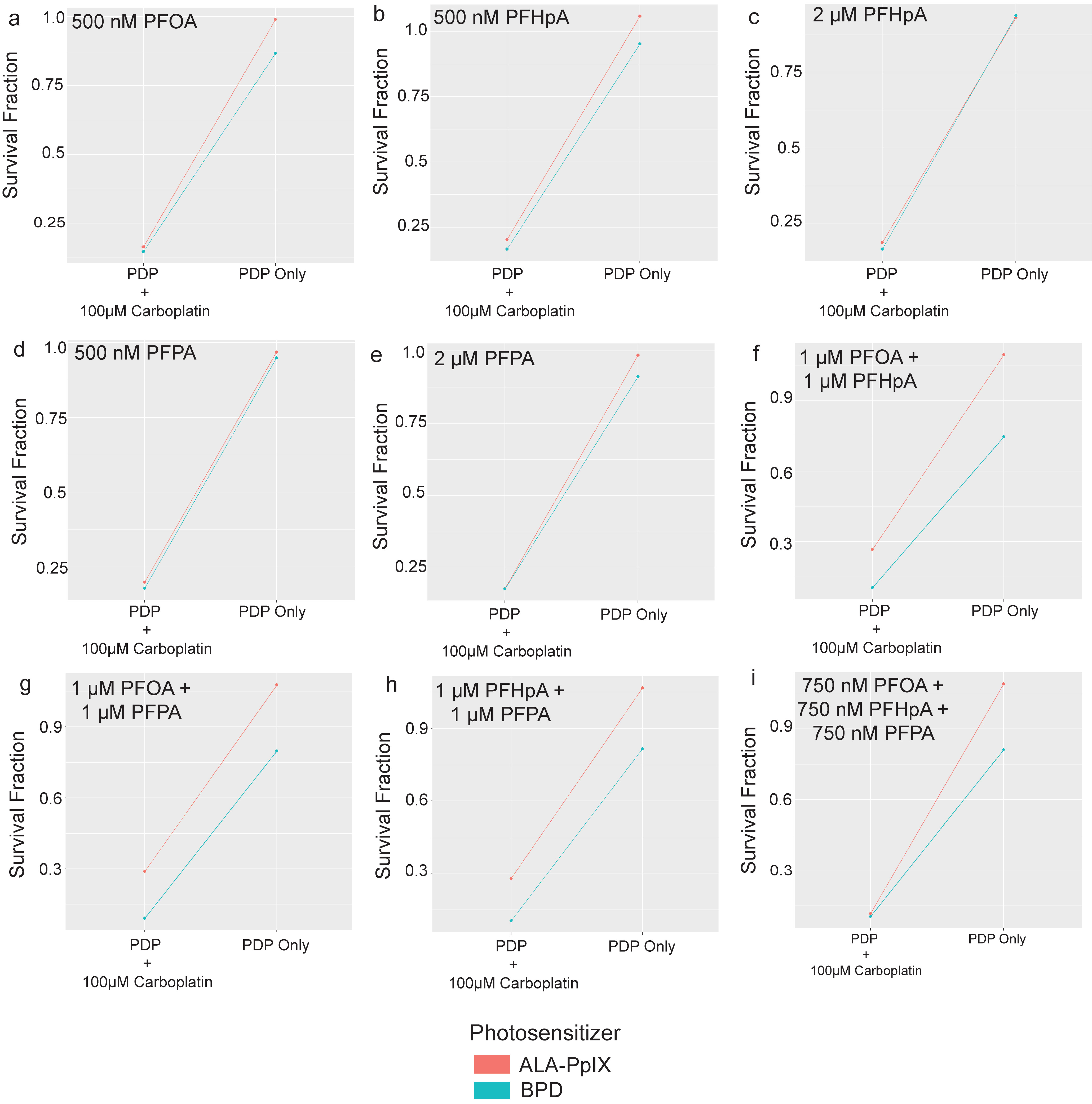
Figure 6. BPD-PDP and ALA-PpIX-PDP overcame resistance to carboplatin induced by PFAS and PFAS mixtures in OVCAR-3 (blue) and Caov-3 (red) cells
- FINAL Figure 6 (512 KB)
- Statistical Analysis 3 (944 KB)
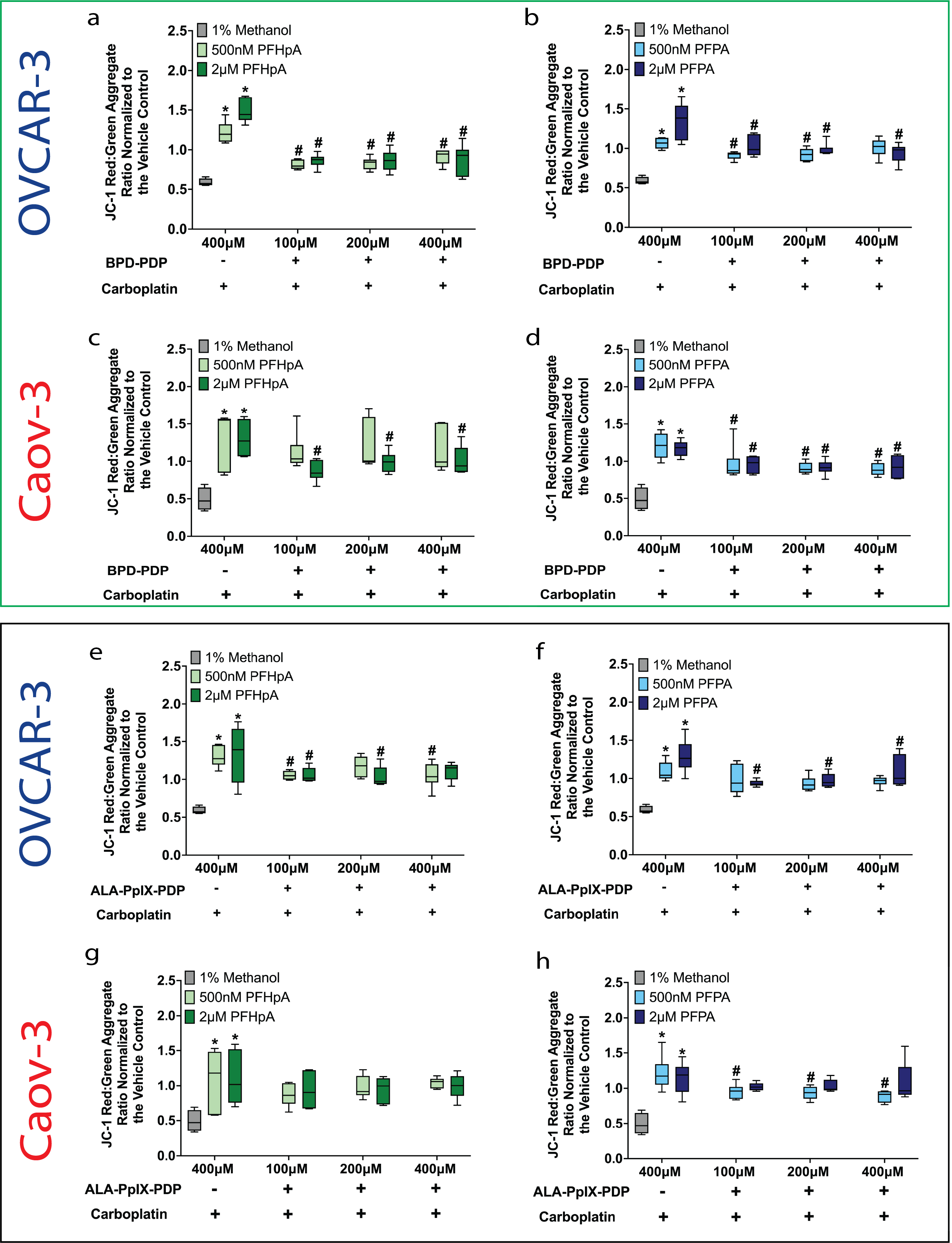
Figure 7. DΨm decreased in PFAS-exposed OVCAR-3 and Caov-3 cells after BPD-PDP (green box) or ALA-PpIX-PDP (black box) in combination with carboplatin
- FINAL Figure 7 (719 KB)
- Statistical Analysis 3 (944 KB)
- Statistical Analysis 4 (902 KB)
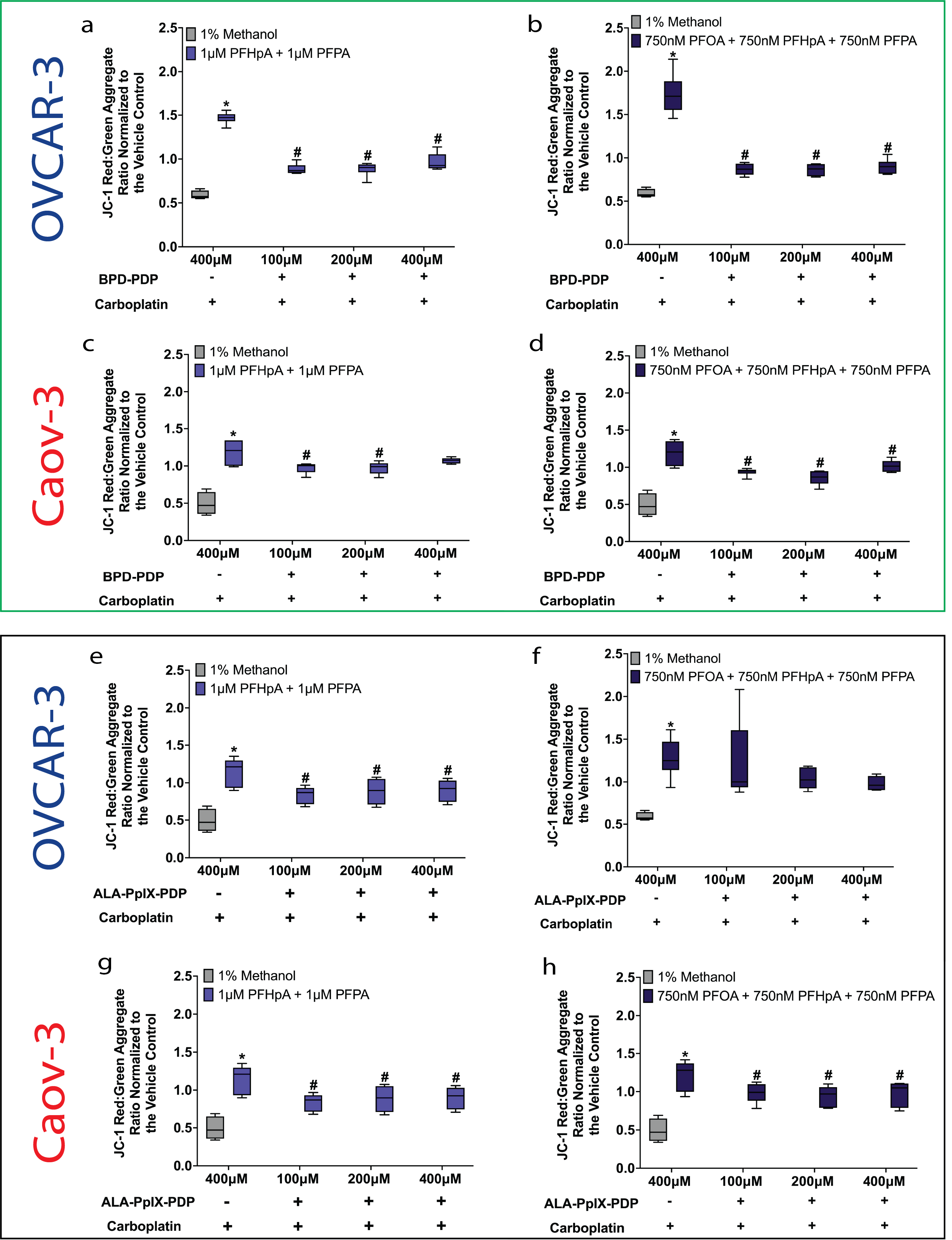
Figure 8. DΨm decreased after BPD-PDP (green box) or ALA-PpIX-PDP (black box) in combination with carboplatin in OVCAR-3 and Caov-3 cells exposed to PFAS mixtures
- FINAL Figure 8 (702 KB)
- Statistical Analysis 3 (944 KB)
- Statistical Analysis 4 (902 KB)
Supplemental Tables
Table S1. Summary of results.
- Table S1 (289 KB)
Supplemental Figures
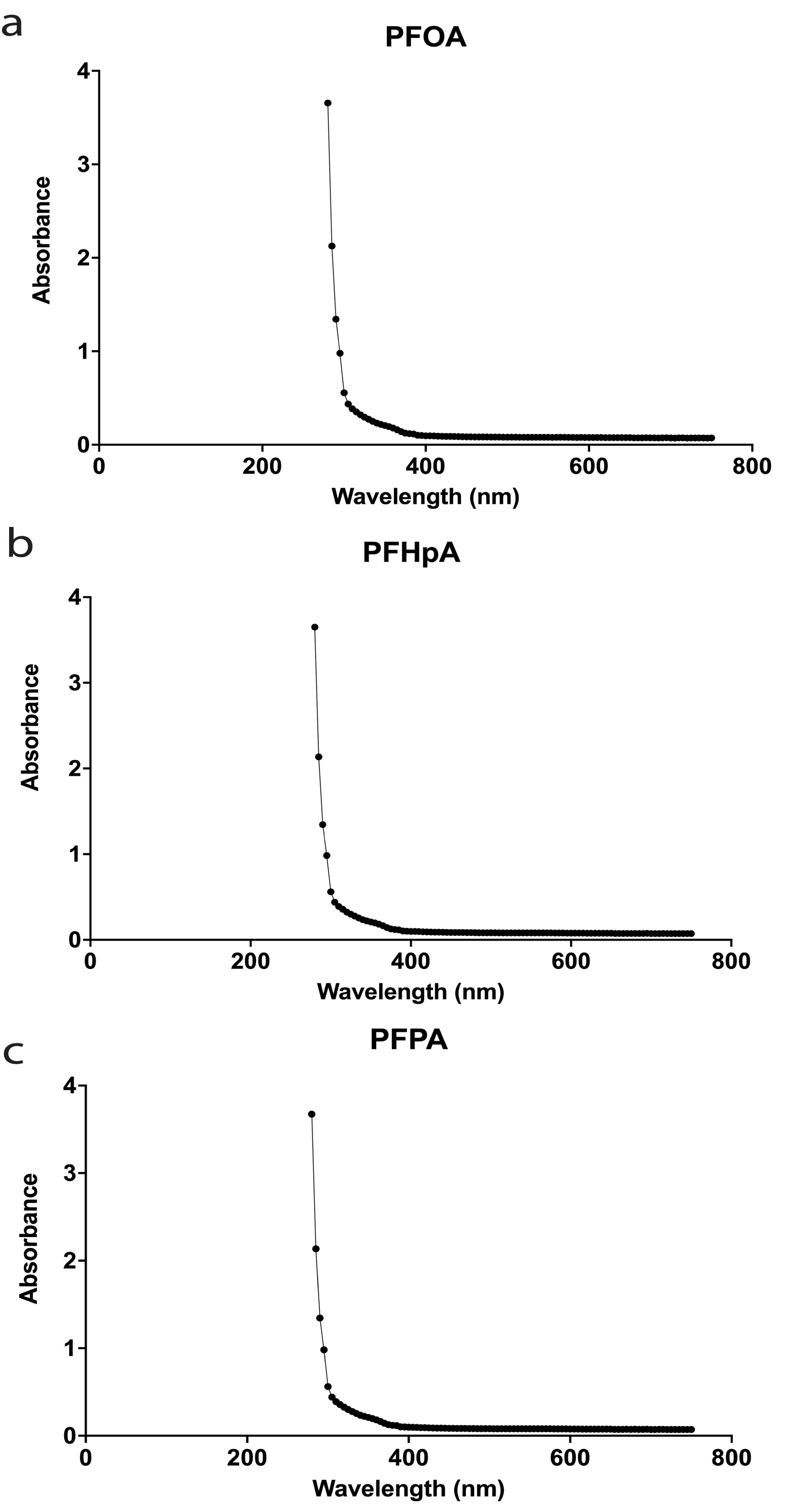
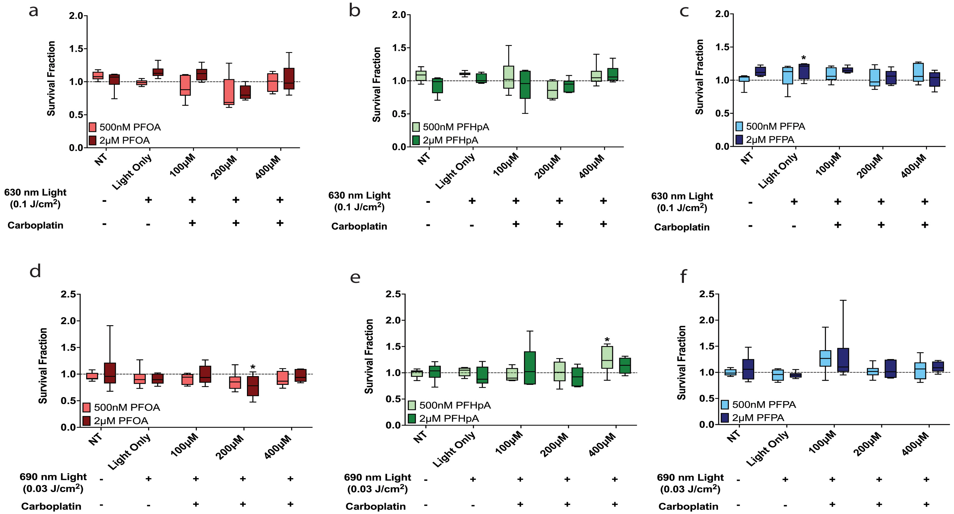
Figure S1. Absorbance spectra for the PFAS agents evaluated in the present study.
- FINAL Figure S1 (167 KB)
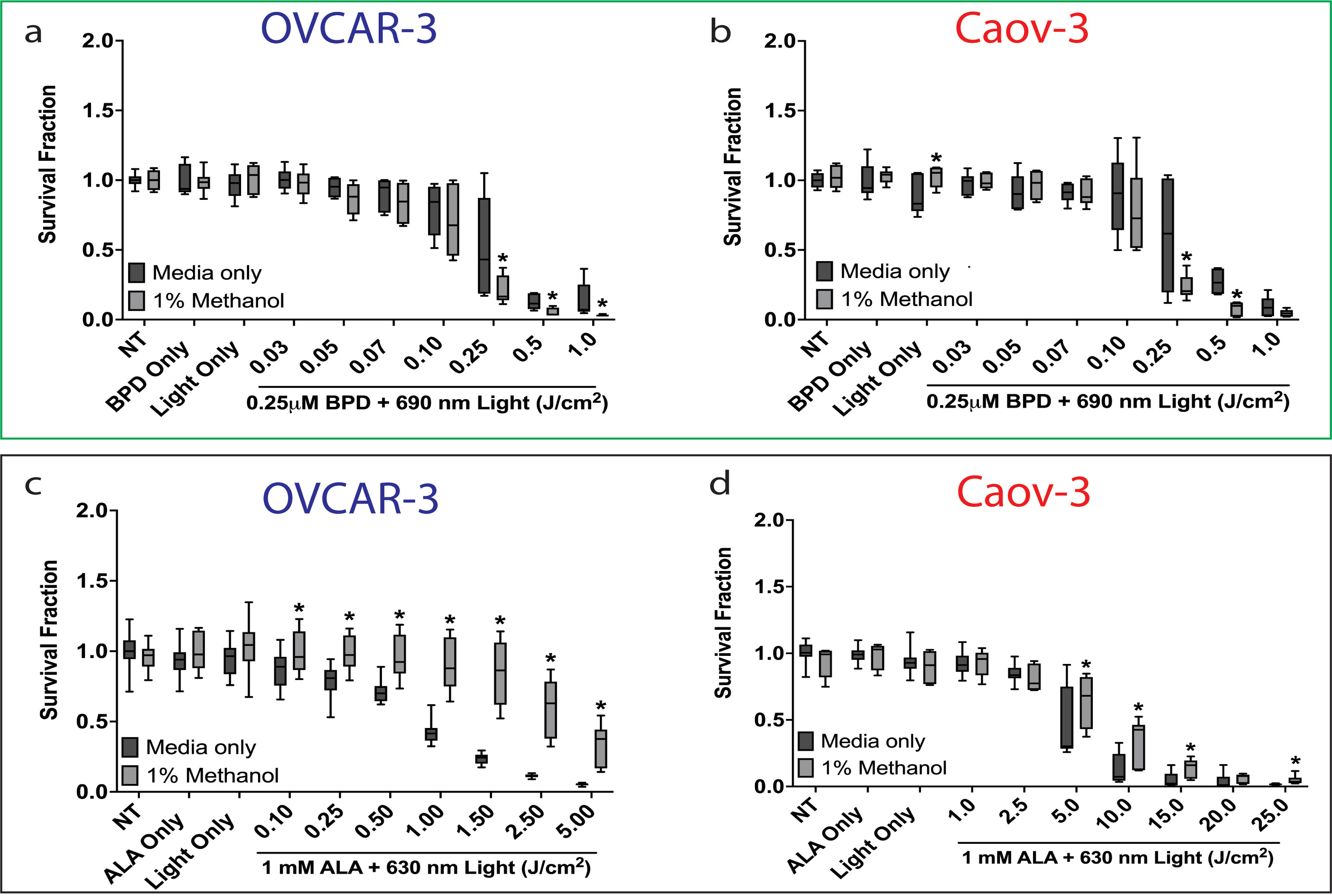
Figure S2. Effect of 1% methanol on survival fraction post-BPD-PDT (green box) or ALA-PpIX-PDT (black box) in OVCAR-3 and Caov-3 cells.
- FINAL Figure S2 (456 KB)
- Statistical Analysis 1 (1 MB)
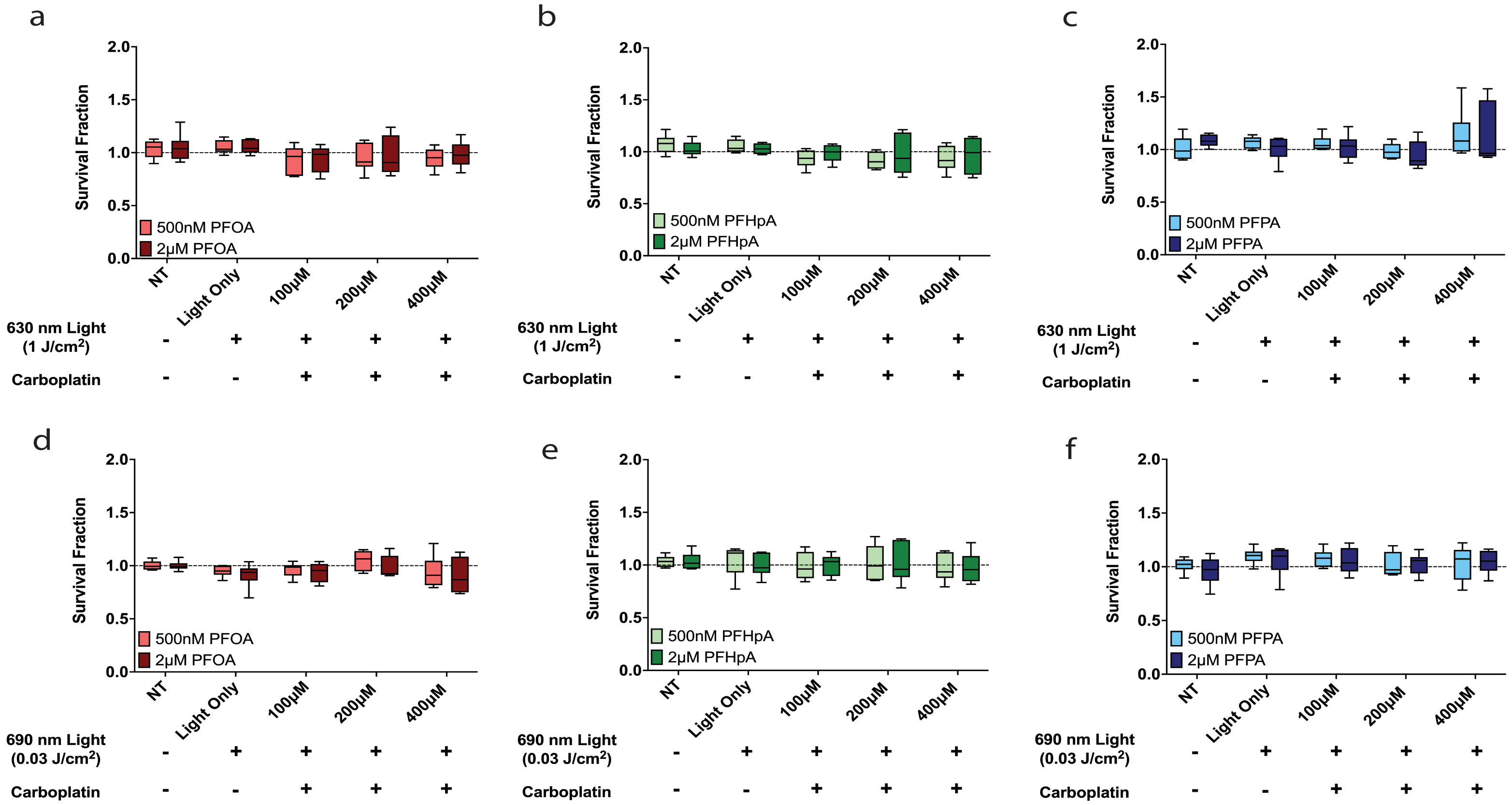
Figure S3. Effect of PFOA on BPD-PDT (green box) and ALA-PpIX-PDT (black box) dose-response curves in OVCAR-3 and Caov-3 cells.
- FINAL Figure S3 (504 KB)
- Statistical Analysis 1 (1 MB)
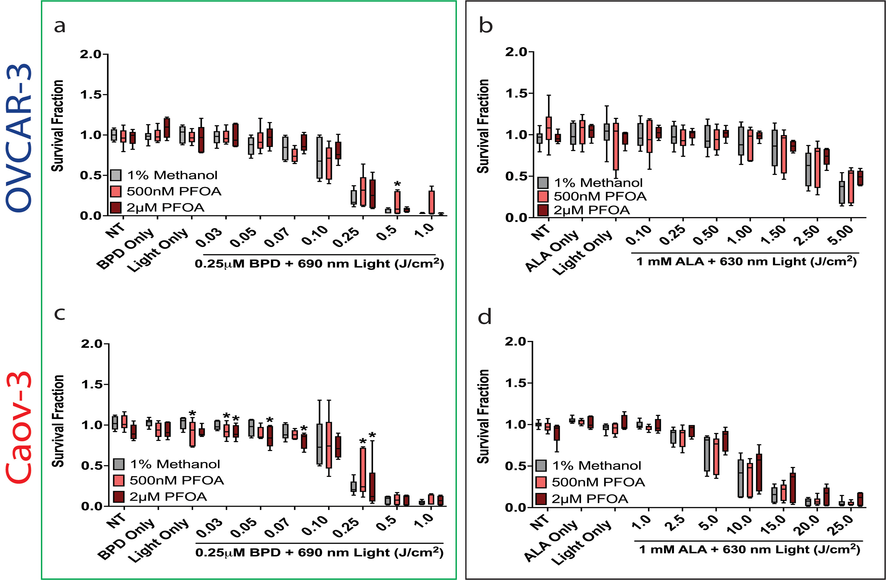
Figure S4. Effect of PFAS mixtures on BPD-PDT (green box) and ALA-PpIX-PDT (black box) dose-response curves in OVCAR-3 and Caov-3 cells.
- FINAL Figure S4 (689 KB)
- Statistical Analysis 1 (1 MB)
Figure S5. Survival fraction decreased in PFAS-exposed OVCAR-3 and Caov-3 cells post-BPD-PDP and ALA-PpIX-PDP + carboplatin.
- FINAL Figure S5 (583 KB)
- Statistical Analysis 2 (1 MB)
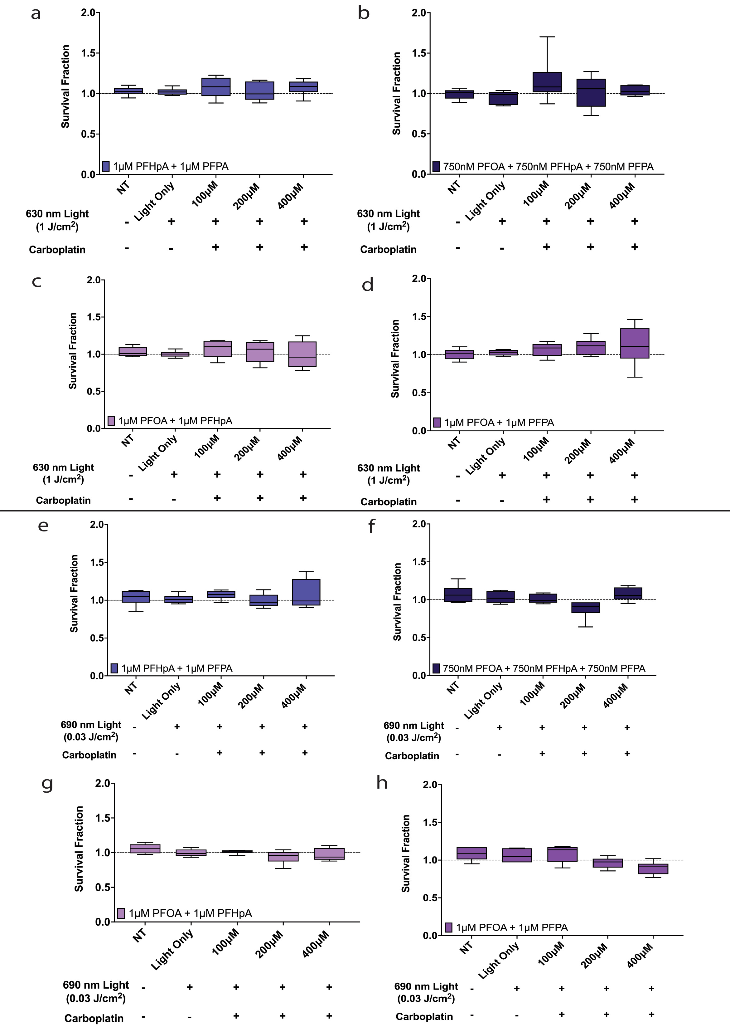
Figure S6. Survival fraction remained largely unchanged in PFAS mixture-exposed OVCAR-3 and Caov-3 cells post-BPD-PDP or ALA-PpIX-PDP.
- FINAL Figure S6 (348 KB)
- Statistical Analysis 2 (1 MB)
Figure S7. In OVCAR-3 cell exposure groups where platinum resistance was observed, photosensitizer efficacy for PDP in combination with 100 μm carboplatin did not differ between BPD and ALA-PpIX
- FINAL Figure S7 (908 KB)
- Statistical Analysis 5 (1 MB)
Figure S8. In Caov-3 cell exposure groups where platinum resistance was observed, photosensitizer efficacy for PDP in combination with 100 μm carboplatin did not differ between BPD and ALA-PpIX.
- FINAL Figure S8 (612 KB)
- Statistical Analysis 5 (1 MB)
Figure S9. Survival fraction in OVCAR-3 cells was largely unaffected by 630 nm or 690 nm light after PFAS exposure.
- FINAL Figure S9 (376 KB)
- Statistical Analysis 2 (1 MB)
Figure S10. Survival fraction in Caov-3 cells was not affected by 630 nm or 690 nm light after PFAS exposure.
- FINAL Figure S10 (360 KB)
- Statistical Analysis 2 (1 MB)
Figure S11. Survival fraction in OVCAR-3 cells was not affected by 630 nm or 690 nm light after exposure to PFAS mixtures.
- FINAL Figure S11 (487 KB)
- Statistical Analysis 2 (1 MB)
Figure S12. Survival fraction in Caov-3 cells was not affected by 630 nm or 690 nm light after exposure to PFAS mixtures.
- FINAL Figure S12 (453 KB)
- Statistical Analysis 2 (1 MB)
Figure S13. ΔΨm decreased in PFOA-exposed OVCAR-3 and Caov-3 cells after BPD-PDP (green box) or ALA-PpIX-PDP (black box) and carboplatin treatment.
- FINAL Figure S13 (423 KB)
- Statistical Analysis 3 (944 KB)
- Statistical Analysis 4 (902 KB)
Figure S14. ΔΨm decreased in PFAS mixture-exposed OVCAR-3 and Caov-3 cells after BPD-PDP (green box) or ALA-PpIX-PDP (black box) and carboplatin treatment.
- FINAL Figure S14 (641 KB)
- Statistical Analysis 3 (944 KB)
- Statistical Analysis 4 (902 KB)
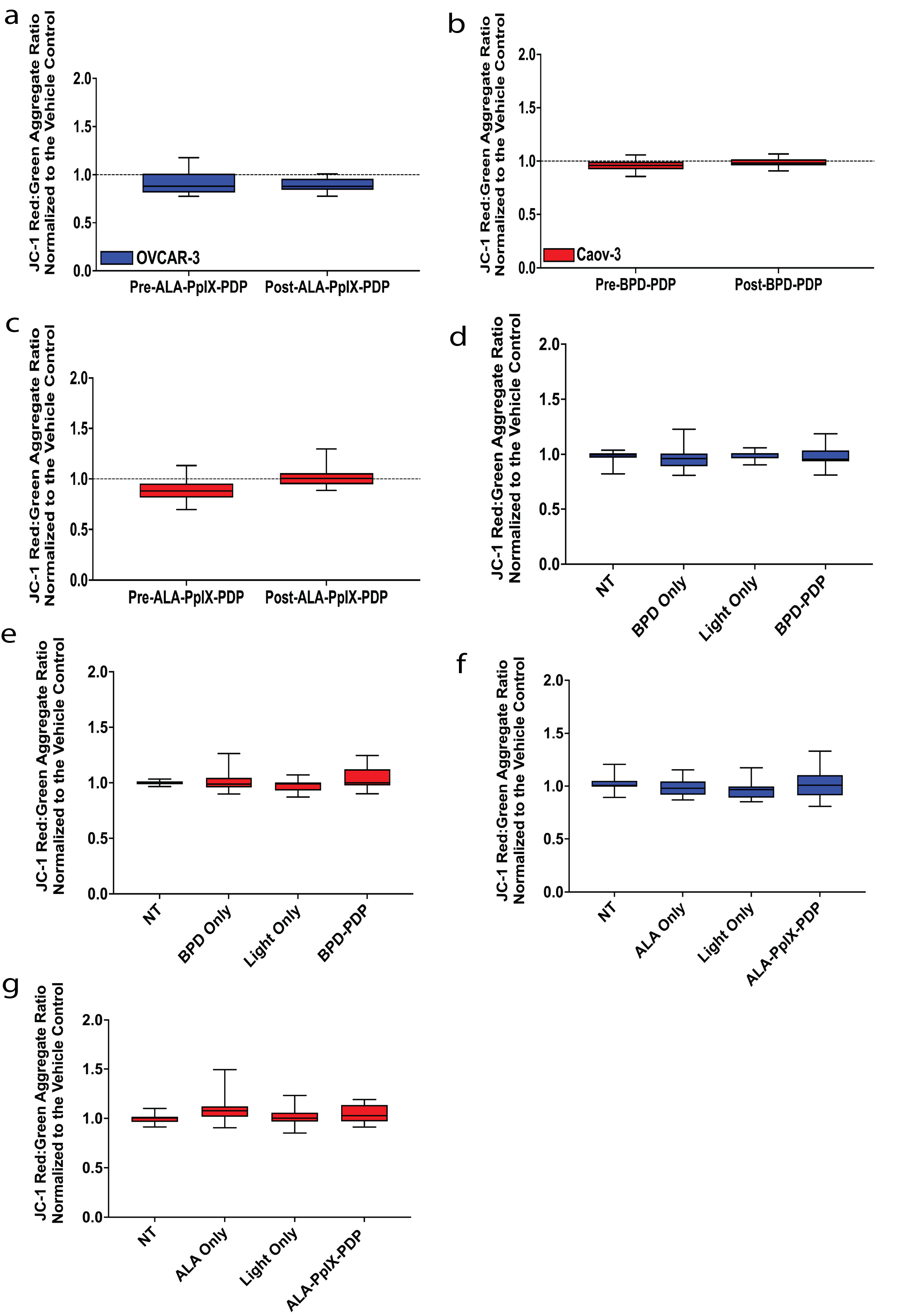
Figure S15. ΔΨm was unchanged in OVCAR-3 (blue) and Caov-3 (red) cells pre- and post-BPD-PDP or ALA-PpIX-PDP, photosensitizer incubation, and irradiation.
- FINAL Figure S15 (443 KB)
- Statistical Analysis 3 (944 KB)
- Statistical Analysis 4 (902 KB)